


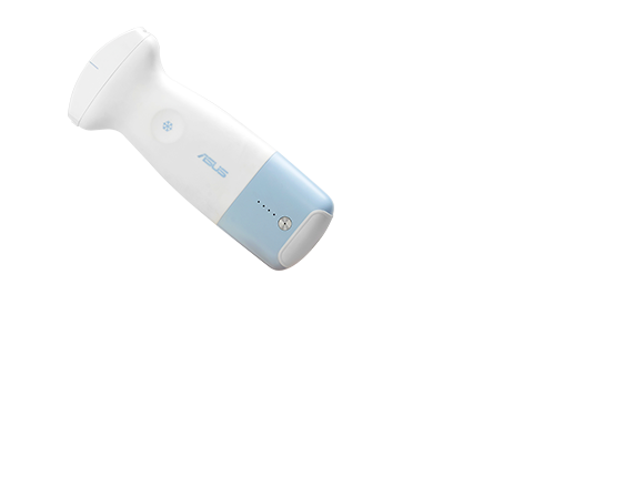
ASUS Handheld Ultrasound Solution LU800
ASUS Handheld Ultrasound Solution LU800
Ultrasound scans,
anytime anywhere.
ASUS Handheld Ultrasound LU800 series
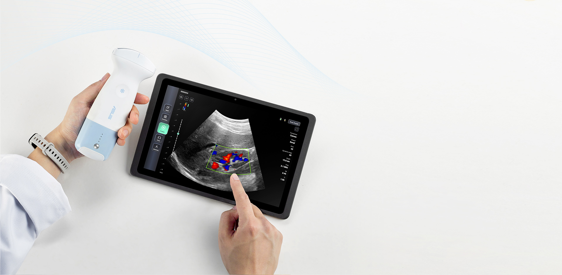
A Point-Of Care Ultrasound system just in your hand
It easily connects to a compatible smartphone or tablet, and is immediately ready for use. This lightweight device is compact and durable, allowing it to be deployed in ambulances, operating theaters, or for training purposes.
-
High quality images
128-channel
beam former
-
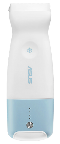
Wireless design
270 g
lightweight
-
With
5
image modes
-
Supports
DICOM
system
-
For all scenarios
IP44
water resistance
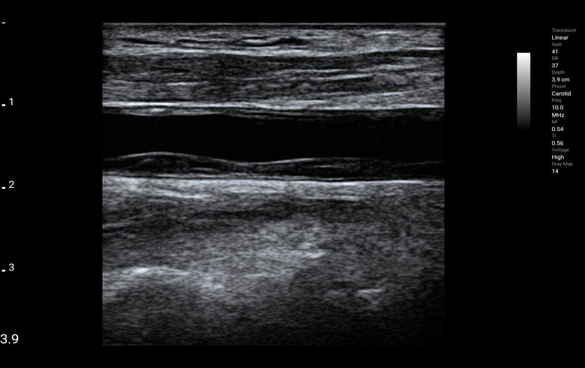
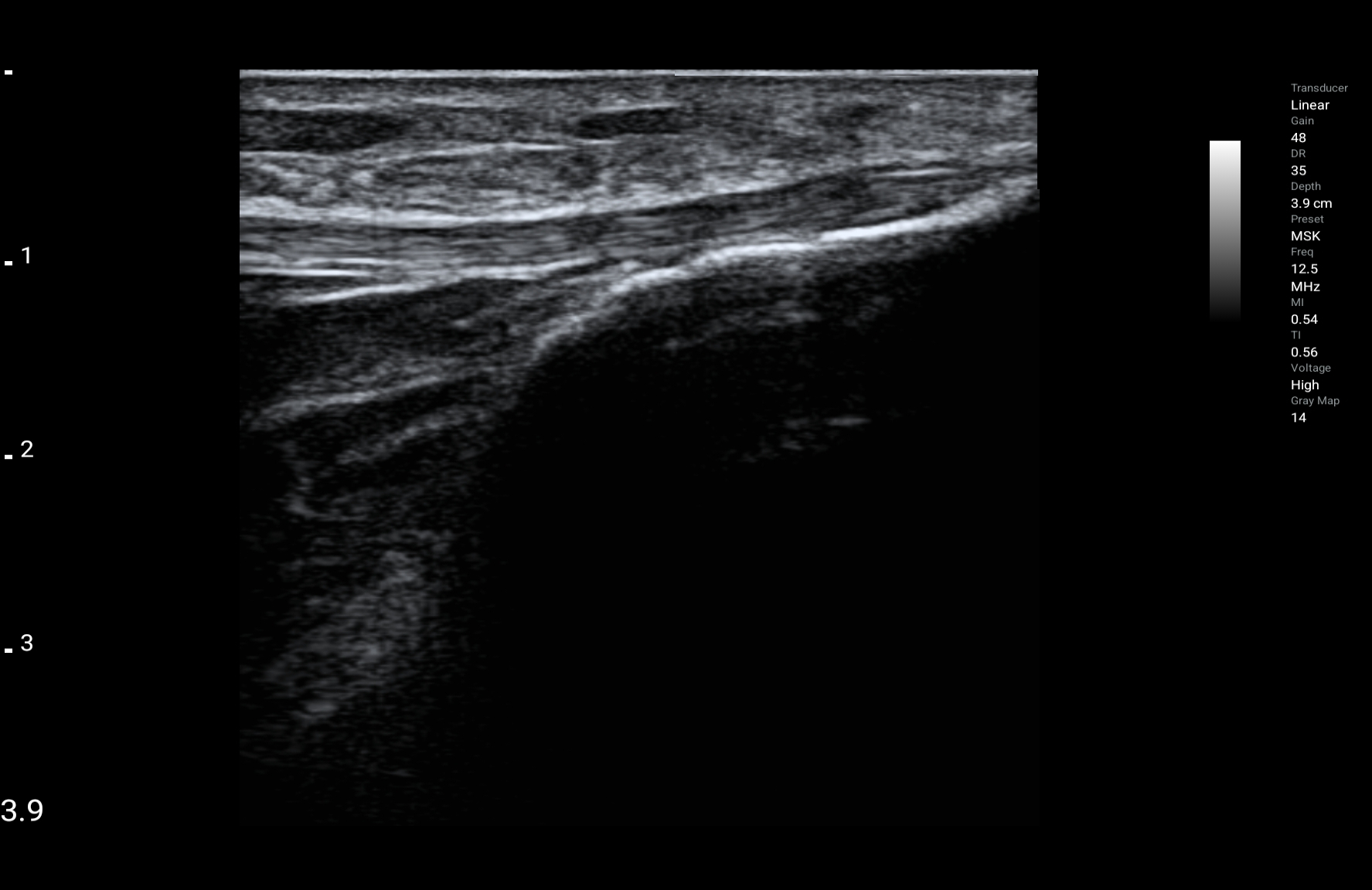
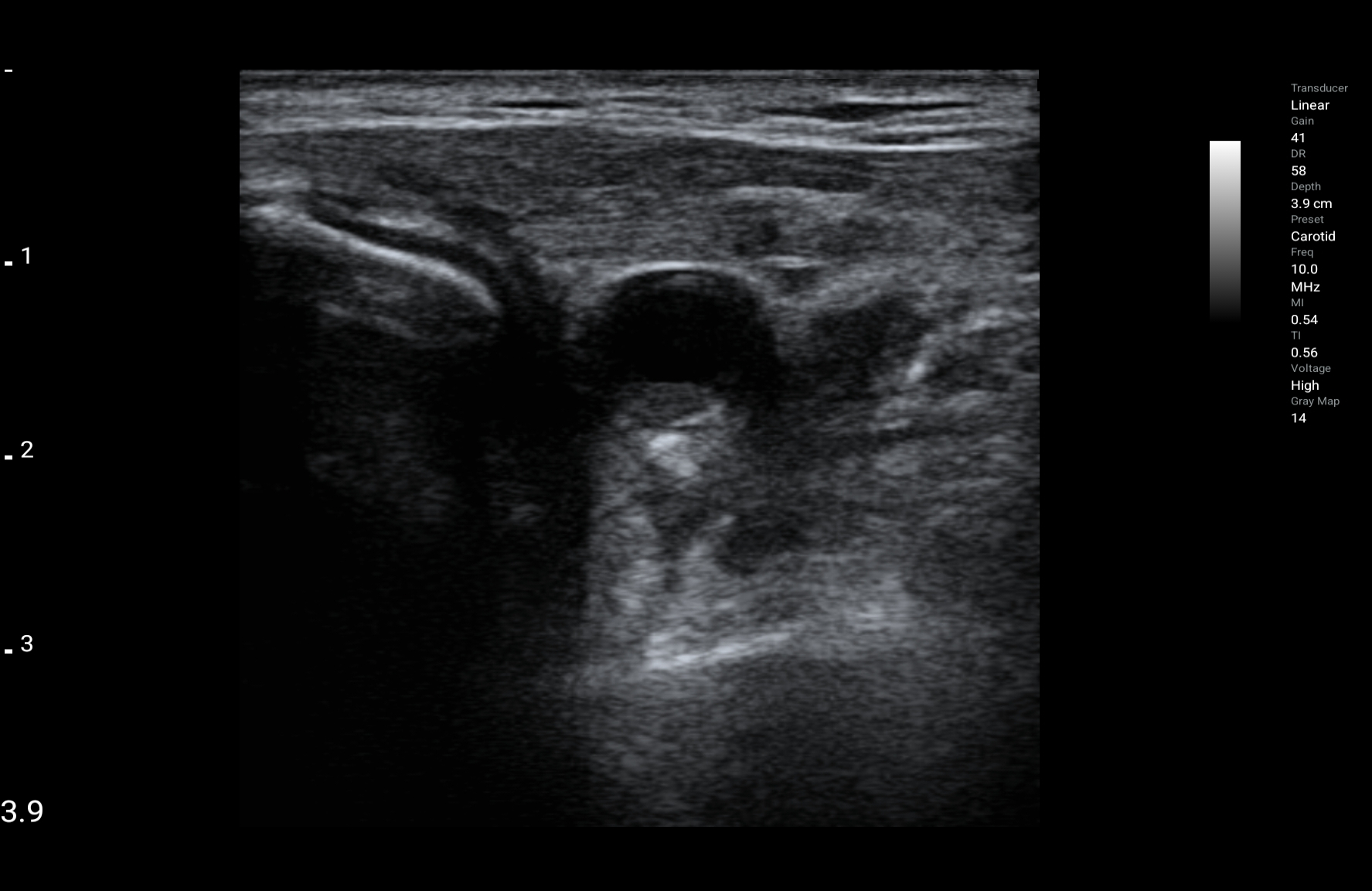
LU800L - Linear
Frequency : 4.2-12.5MHz
Max depth: 12.6cm
-

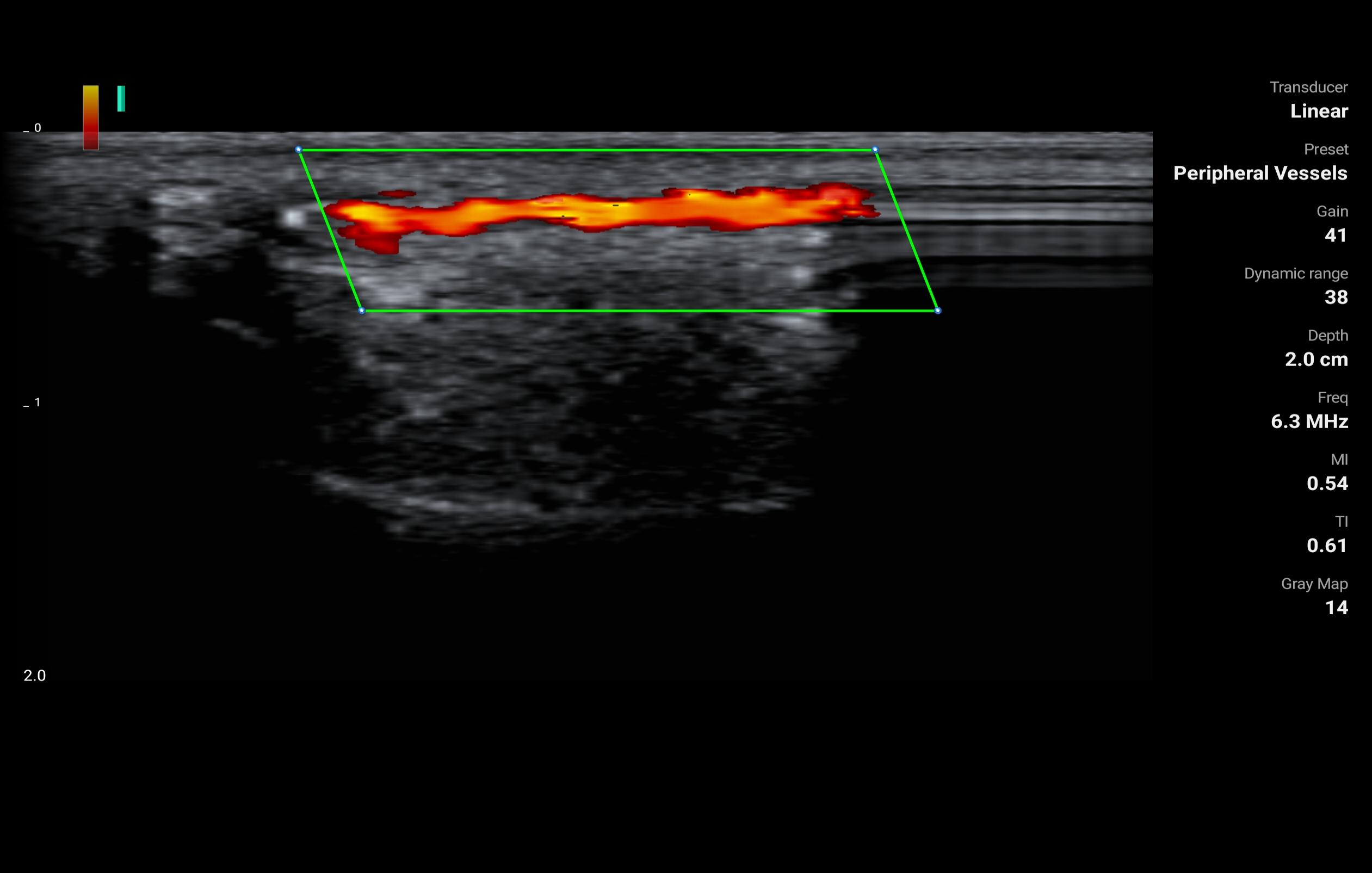
Peripheral vessels
-

Thyroid
-

Breast
-

Superficial
-

Musculoskeletal
-

Carotid
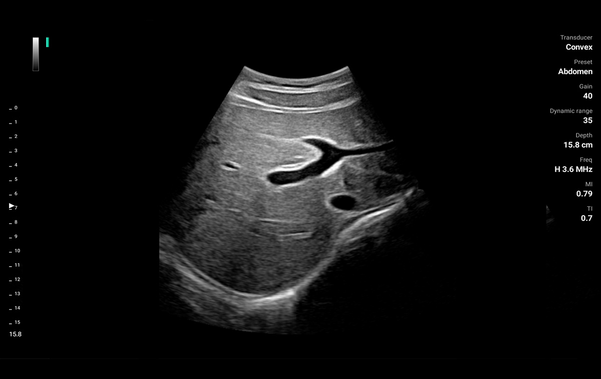
LU800C - Convex
Frequency: 2-5MHz
Max depth: 30cm
Field of view: 60°
-

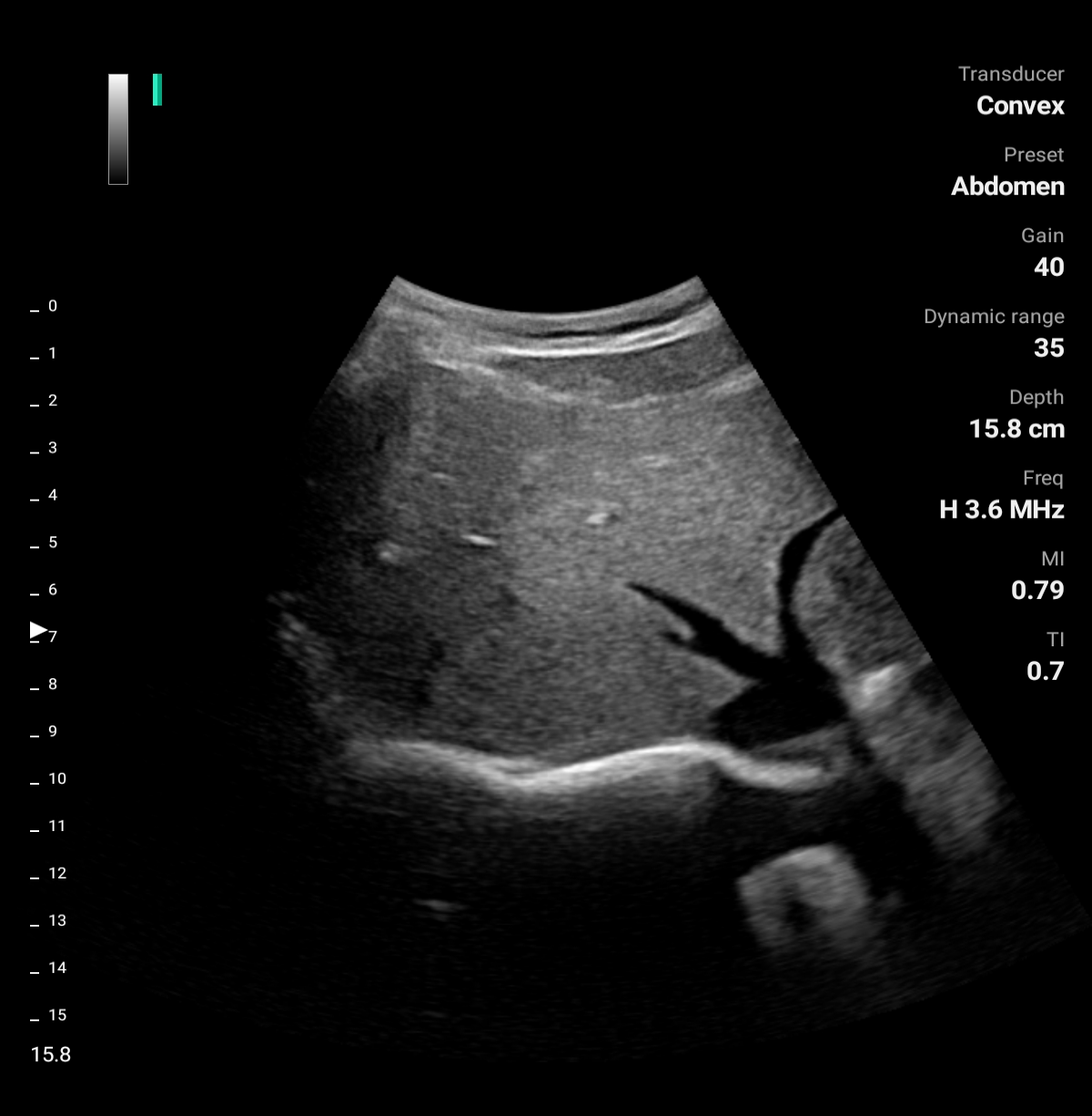
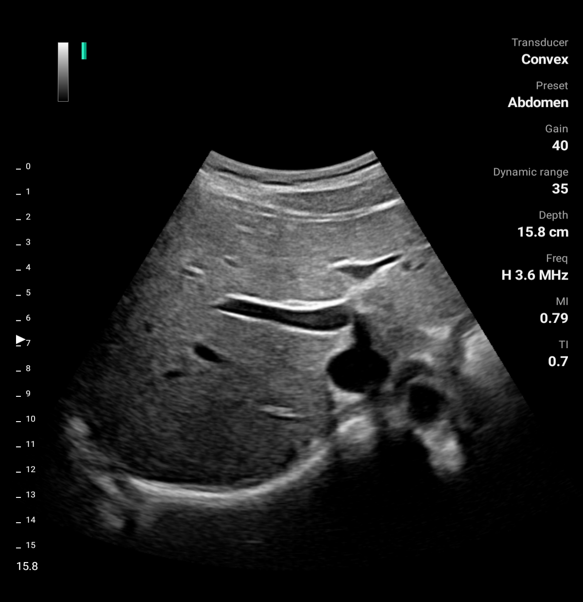
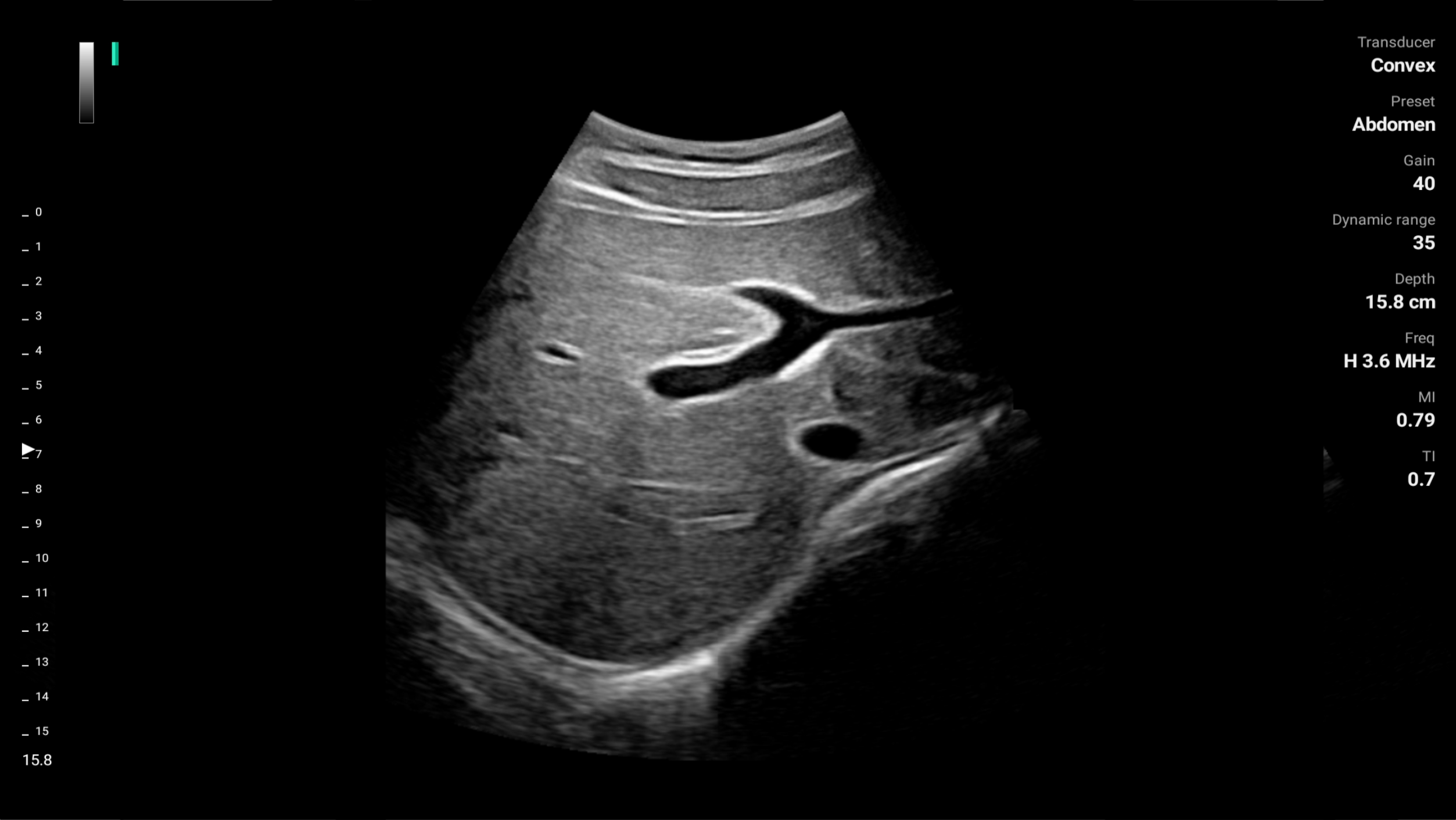
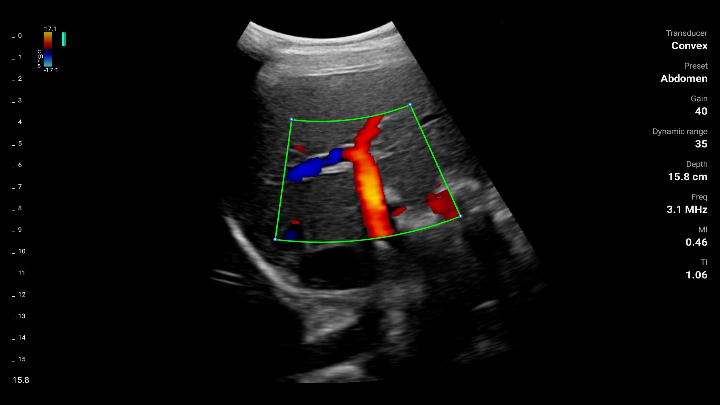
Abdomen
-

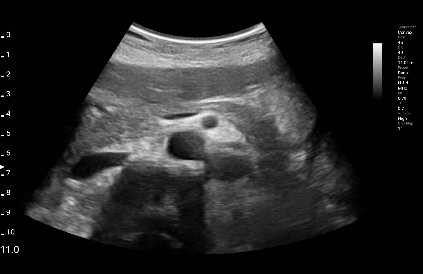
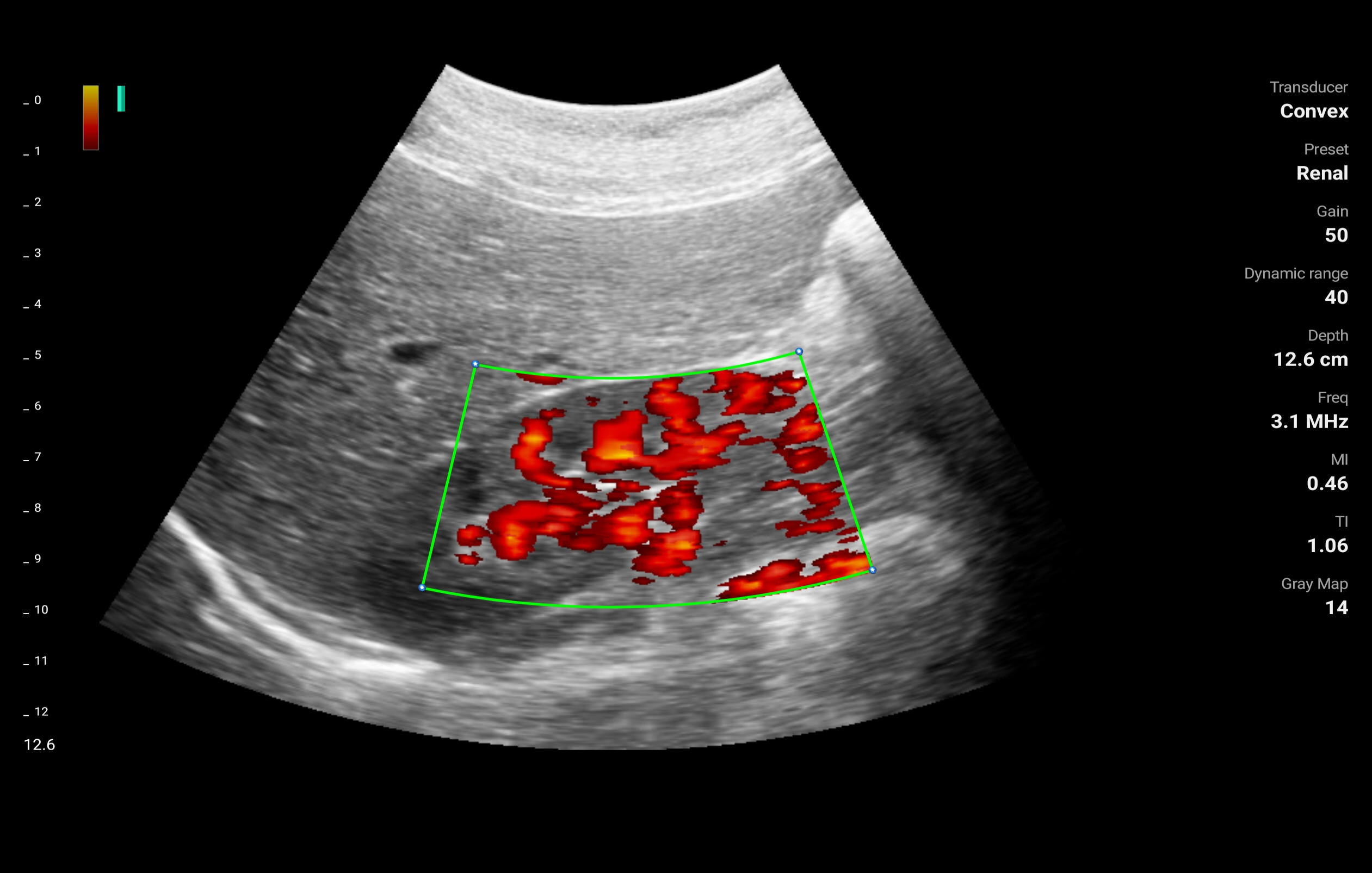
Renal
-

GYN
-

OB Mid Late
-

Bladder Meas
-

FAST
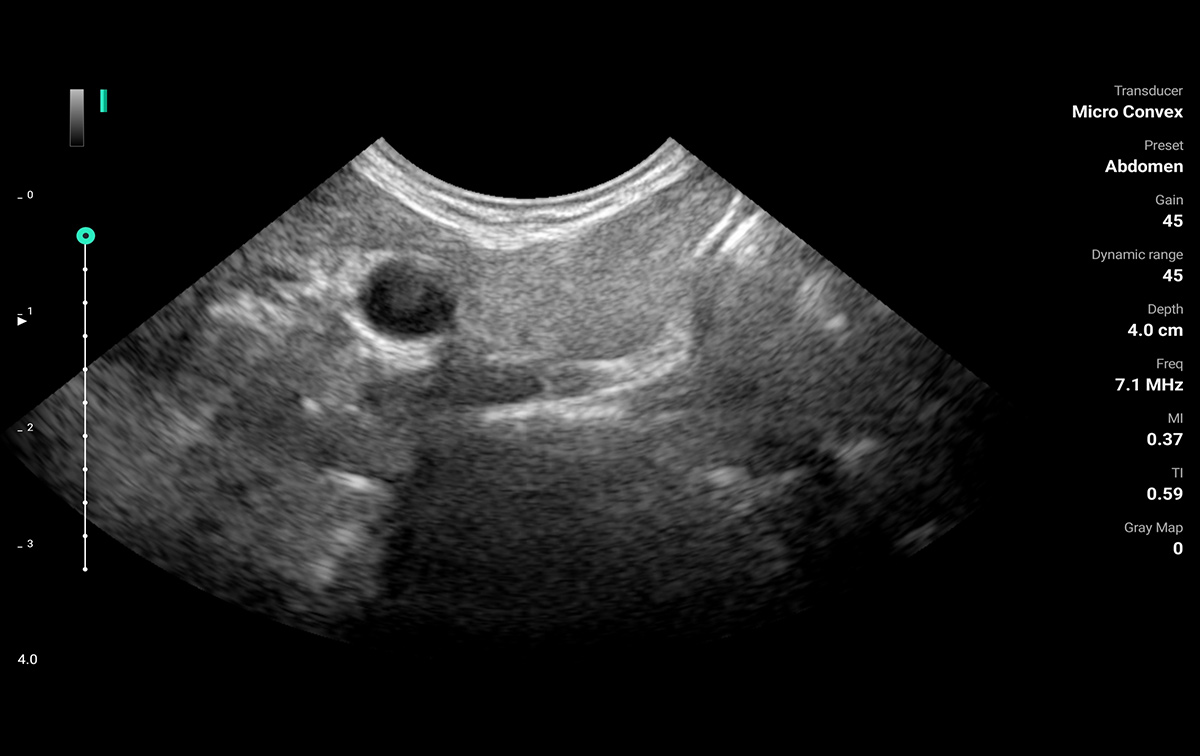
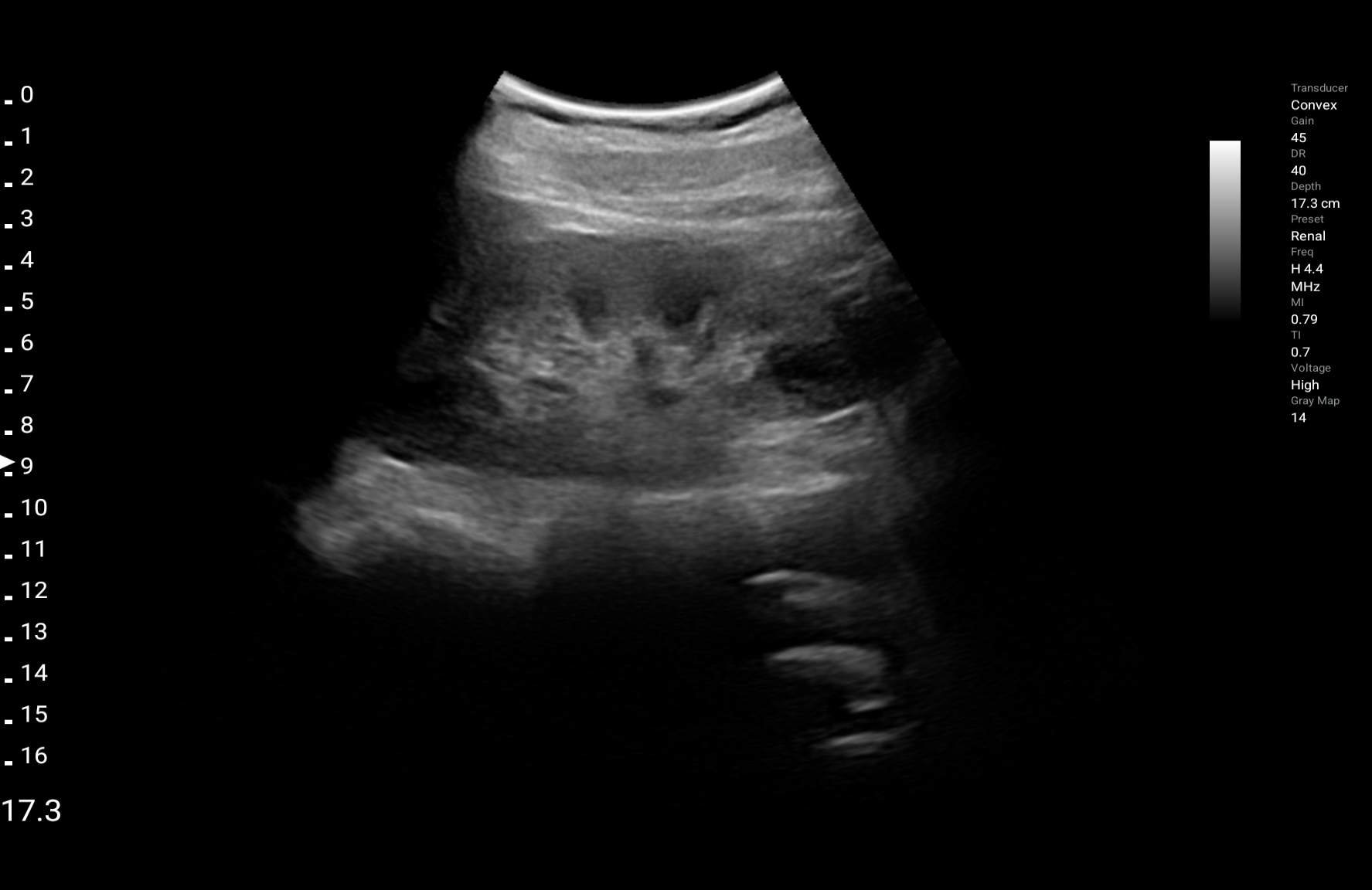
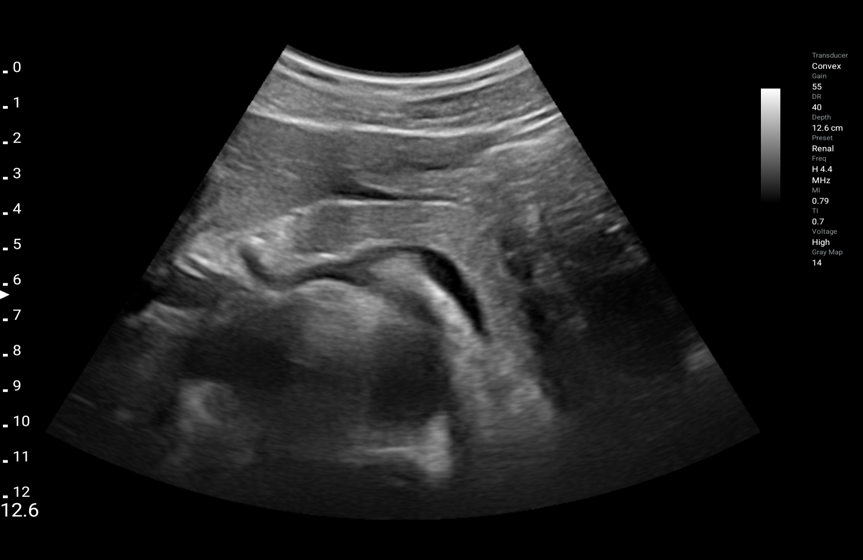
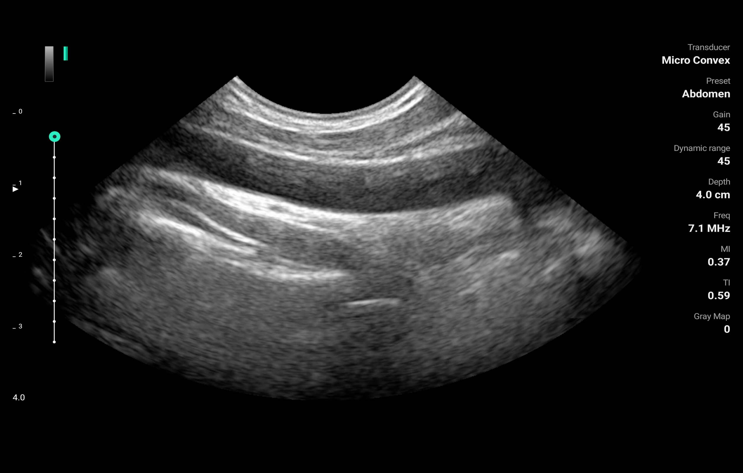
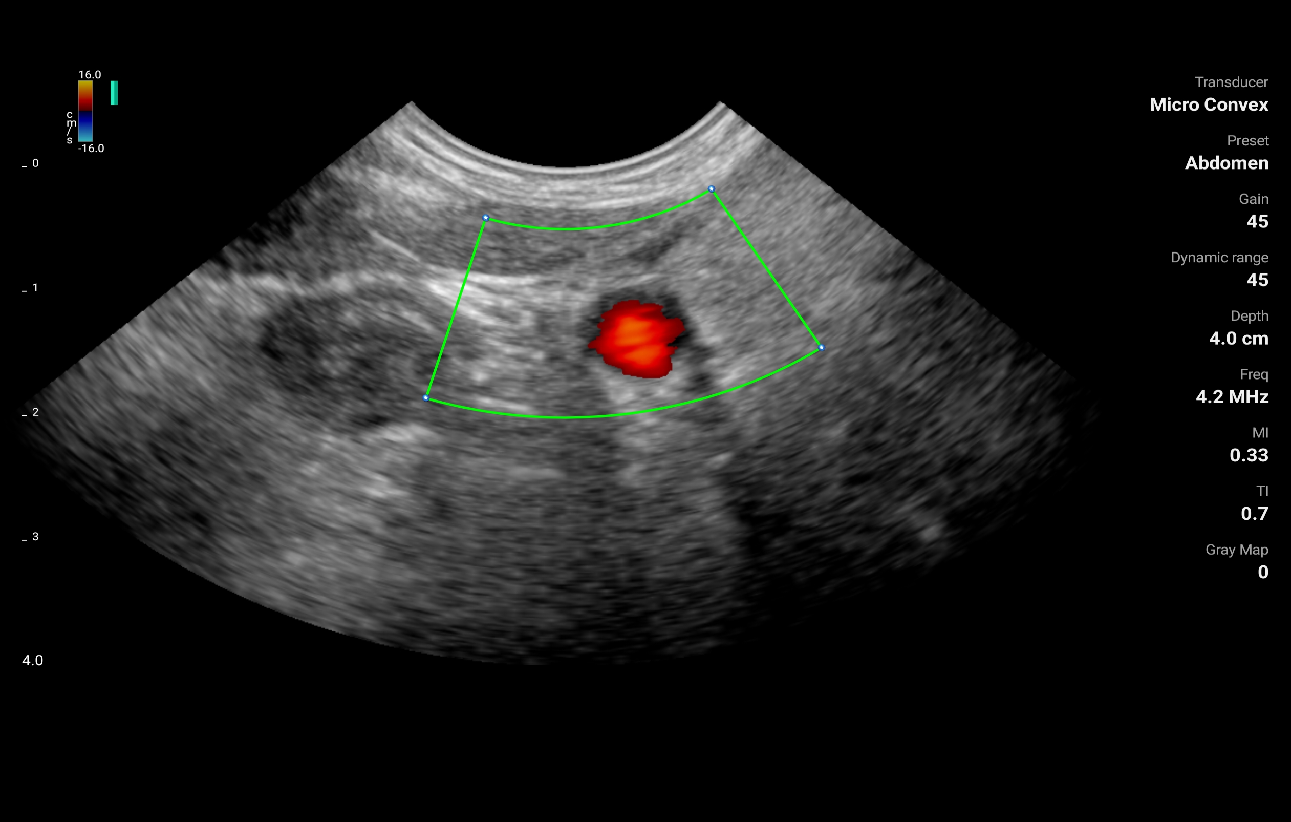
LU800M - Microconvex
Frequency: 3.6-8.5MHz
Max depth: 12.6cm
Field of view: 100°
-
Abdomen
LU800PA - Phased Array
Frequency: 1.3-3.7MHz
Max depth: 30cm
Field of view: 90°
-
Abdomen
-

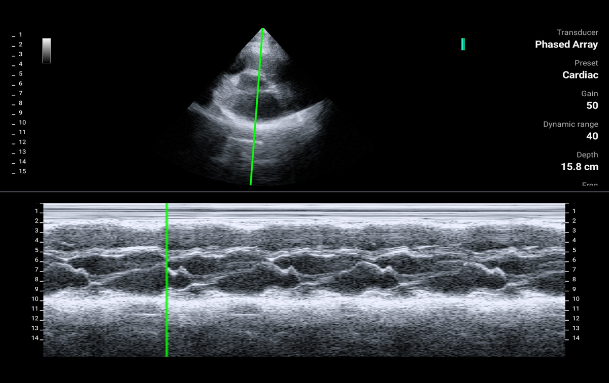
Cardiac
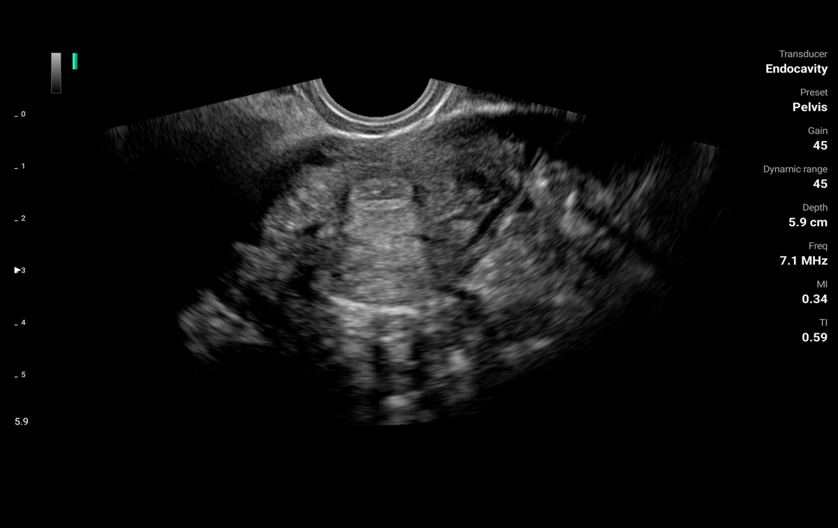
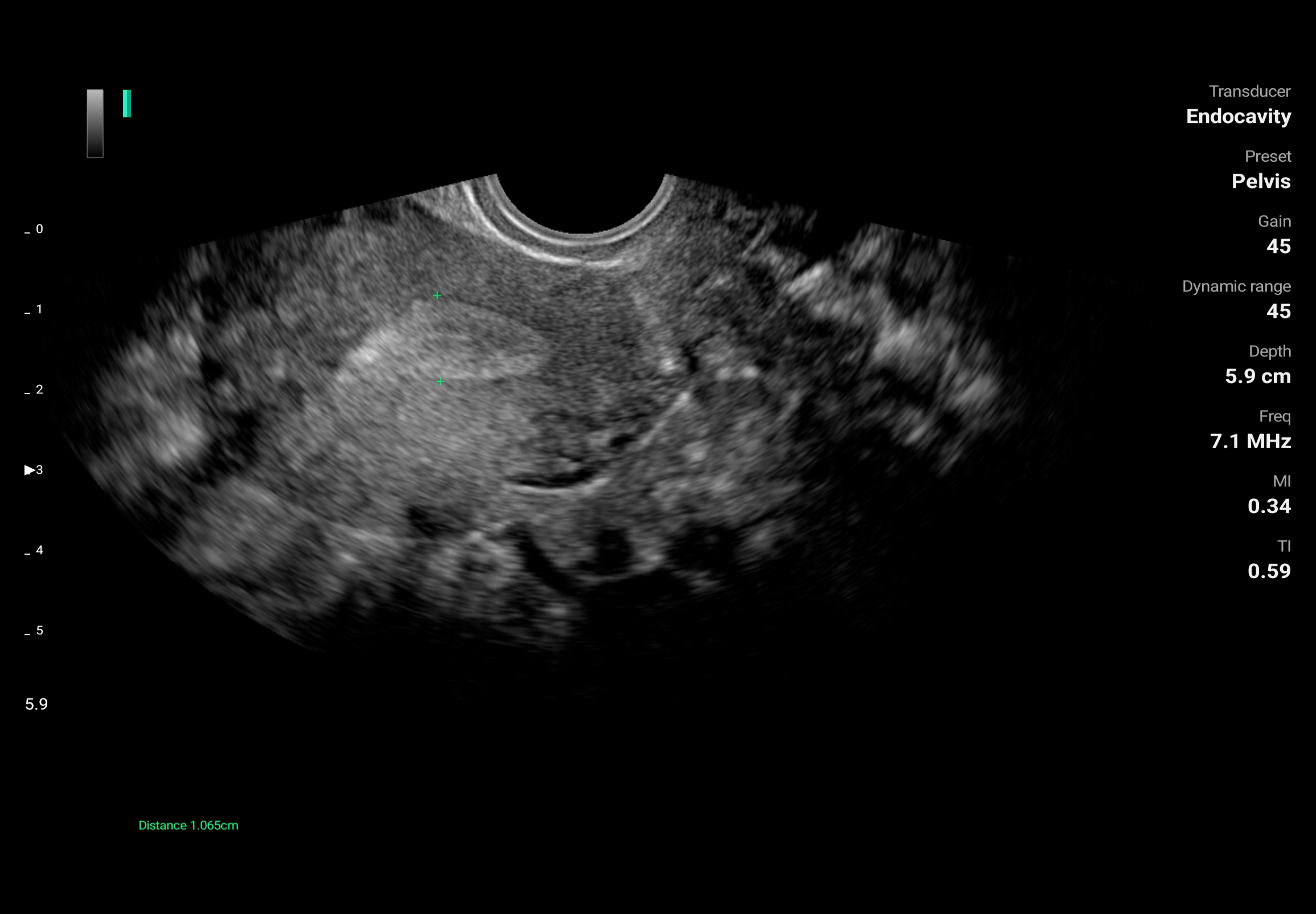
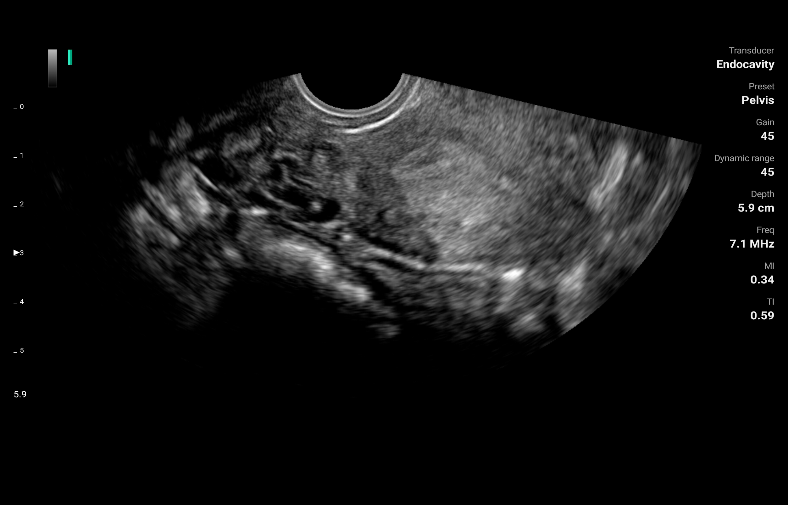
LU800E - Endocavity
Frequency: 3.6-8.5MHz
Max depth: 15cm
Field of view: 151°
-

Pelvis
-

Urology
The Pioneer in Wireless Ultrasound Technology
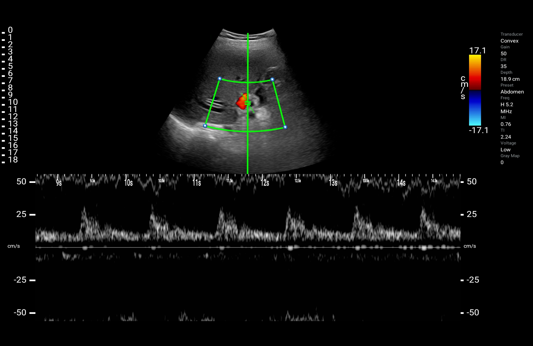
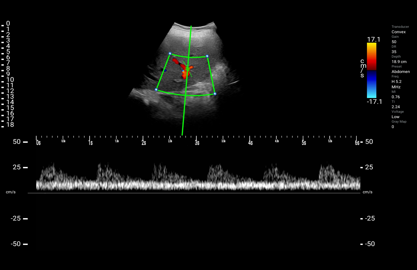
High-quality imagery
ASUS Handheld Ultrasound Solution LU800 features a 128-channel beamformer for clear and sharp ultrasound imagery.

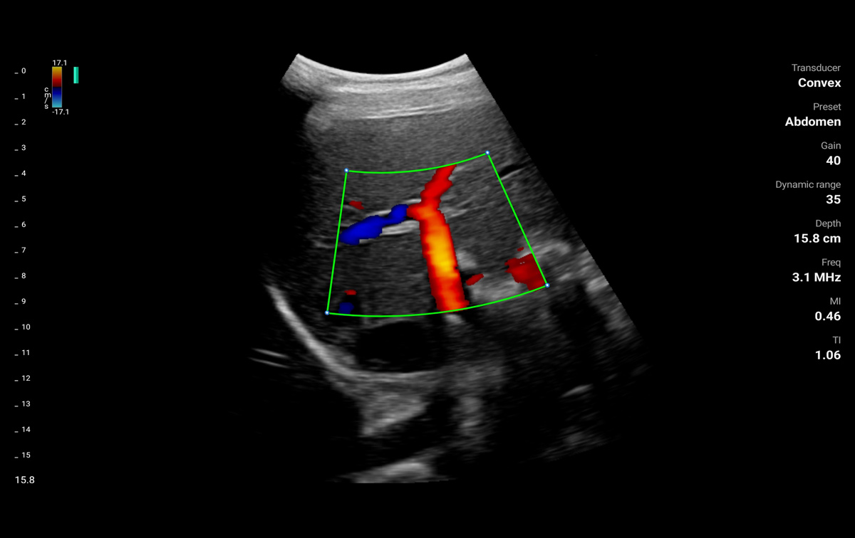
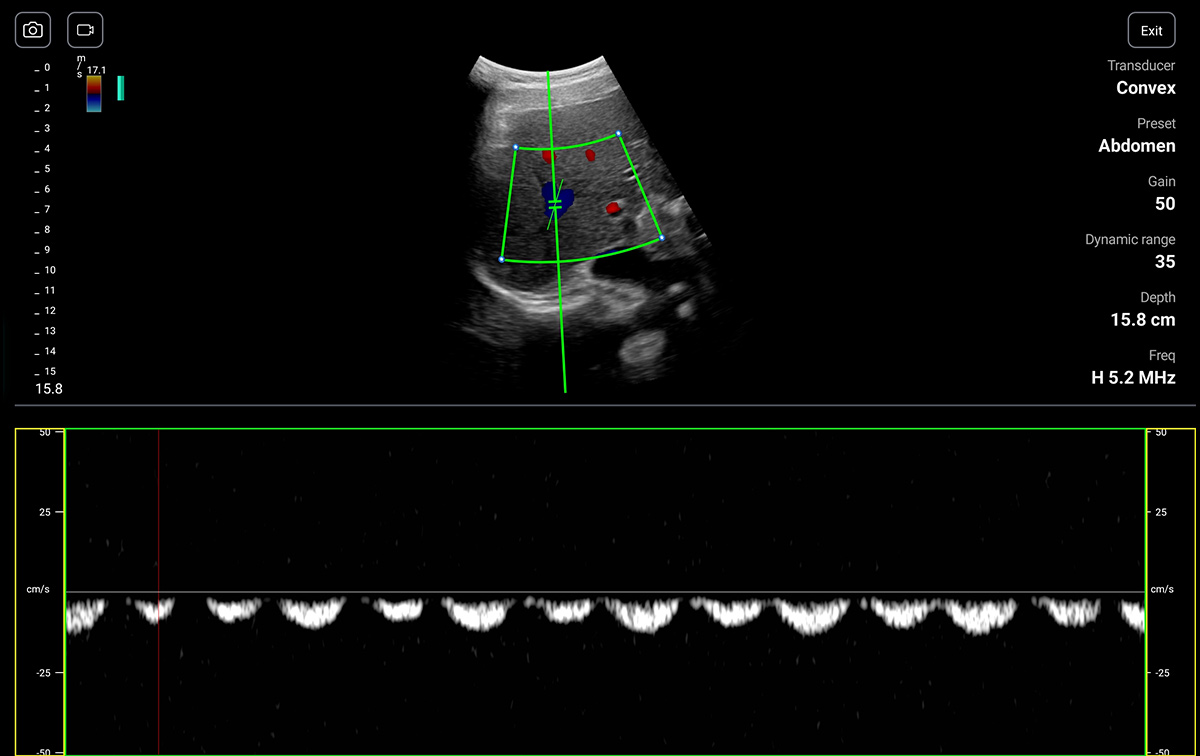
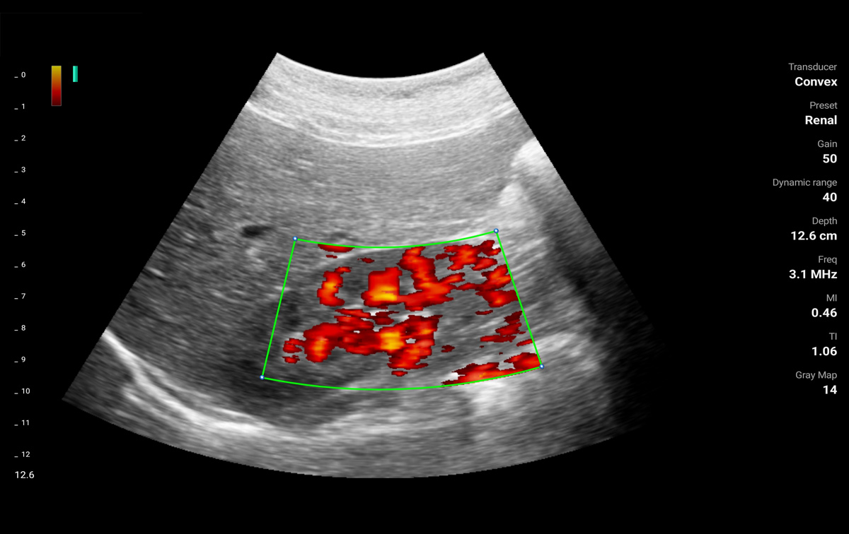
Superior imaging with various scanning modes
Various scanning modes enable clinician for accurate diagnostics and evaluation.
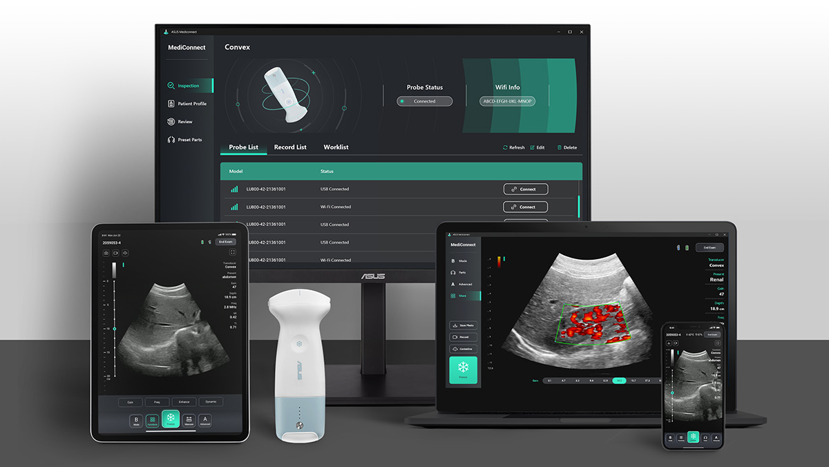
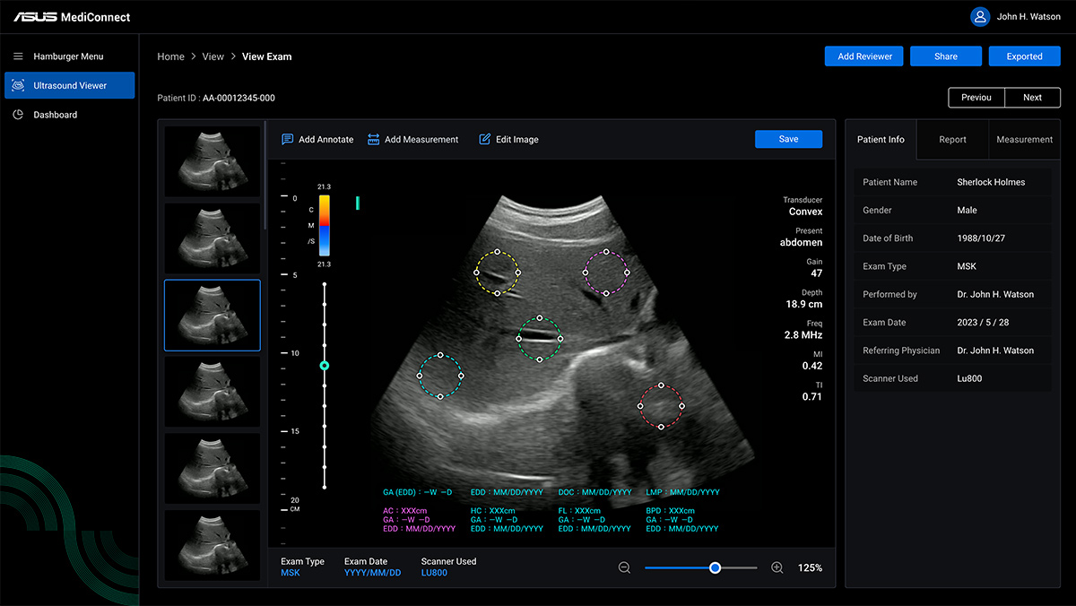
ASUS MediConnect App
Compatible with all mobile devices
*Support DICOM & PACS System


Accelerating Diagnostic Processes with AI-enabled Technology
Powerful functions & tools
Its intuitive interface, advanced tuning function and measurement tools, allows clinician to enhance image clarity and diagnostic accuracy. Leading to more accurate assessments and informed clinical decisions.
Tuning Functions:
Depth
Frequency
Gain
Persistence
Enhancement
TGC
Dynamic range
Gray map
Freeze timer
Color PRF
Color sensitivity
Color angle
Measurement tools:
Bladder measurement
OB early
OB mid-late
Superficial mirror
Cardiac
MSK
AI-enabled
Utilizing AI algorithms and deep learning techniques to analyze ultrasound images in real time. Speed up your diagnostic process and faster treatment planning.
Intima-media thickness (IMT) for carotid
Post-void residual volume (PVR) for bladder urine amount
One-key image optimization
Cloud Viewer
Smart and efficient image management.
Cloud image library
Uploads and downloads
Online image measurement
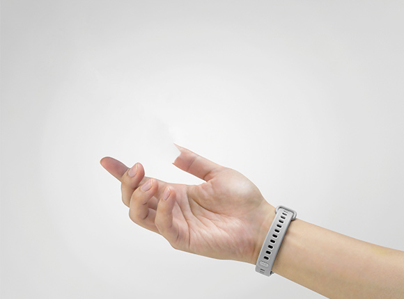
Voice Control : Hands-free imaging
ASUS Handheld Ultrasound Solution LU800 features AI-powered voice commands to provide users with hands-free access to main imaging functions.
Just say it:
Function
Capture Image
Capture Video
Increase Depth
Decrease Depth
Increase Gain
Decrease Gain
Freeze
Live
Center Line
Needle Enhance
Mode Switch
and more …-
Heapatic artery_pw mode
-
.jpg)
Heapatic artery+portal vein PWmode(1)
-
Heapatic artery+portal vein PWmode
-

Heapatic vein PWmode
-
Hepatic vein_B mode
-

Liver Bmode
-
.png)
Liver-Kidney_B mode(1)
-
Liver-Kidney_B mode
-

Liver-Kidney_B mode
-
.png)
Liver-Main portal vein Bmode(1)
-
Liver-Main portal vein Bmode
-
pancreas with heapatic and splenic aryery_B mode
-
Pancreas_B mode
-

Pancreas_B mode
-
Portal vain_B mode
-
Portal vein_CF mode
-
Kidney PD mode
-
.png)
Kidney PD mode (1)
-
.png)
Kidney PD mode (2)
more
close
-
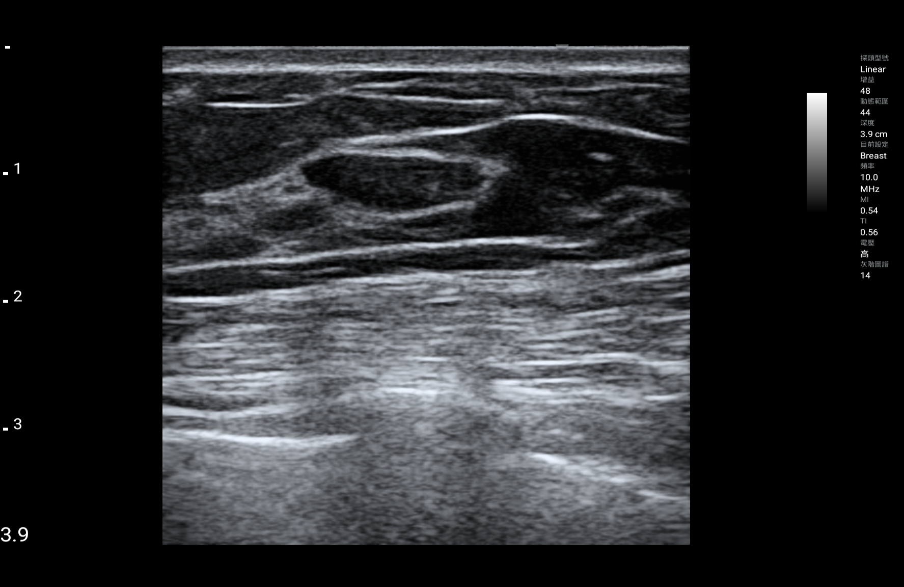
Breast Bmode
-
.png)
Breast Bmode(1)
-
.png)
Breast Bmode(2)
-

Breast cyst Bmode
-
.png)
Apical 4-chamber view_B mode(1)
-
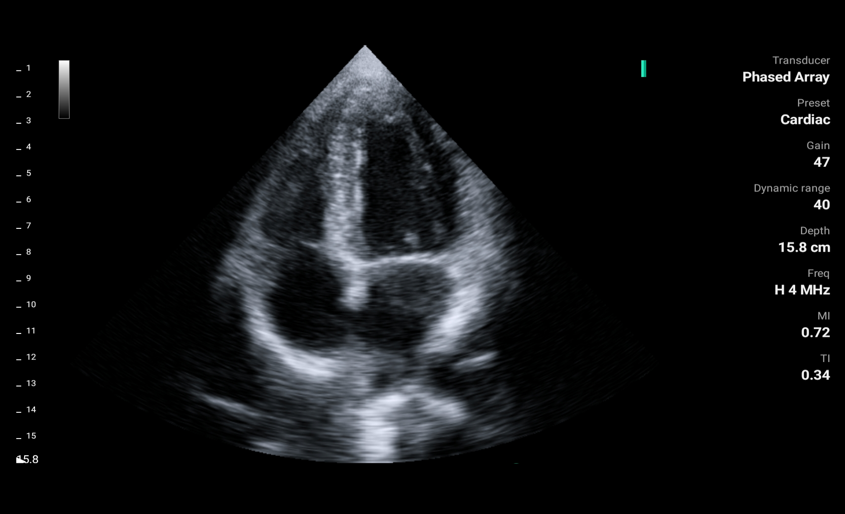
Apical 4-chamber view_B mode
-
.png)
Parasternal long-axis view_B mode(1)
-
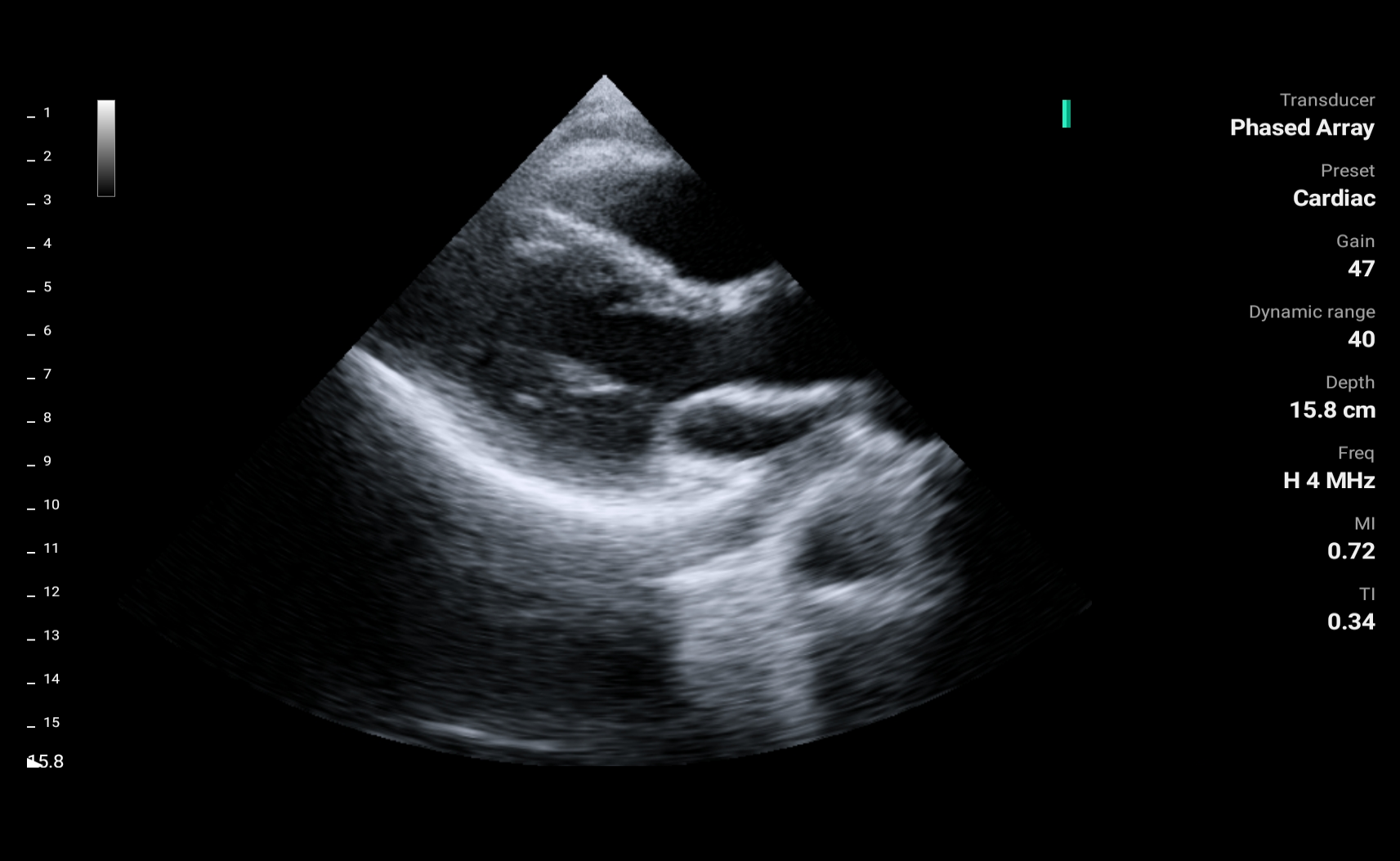
Parasternal long-axis view_B mode
-
.png)
Parasternal long-axis view_M mode(1)
-
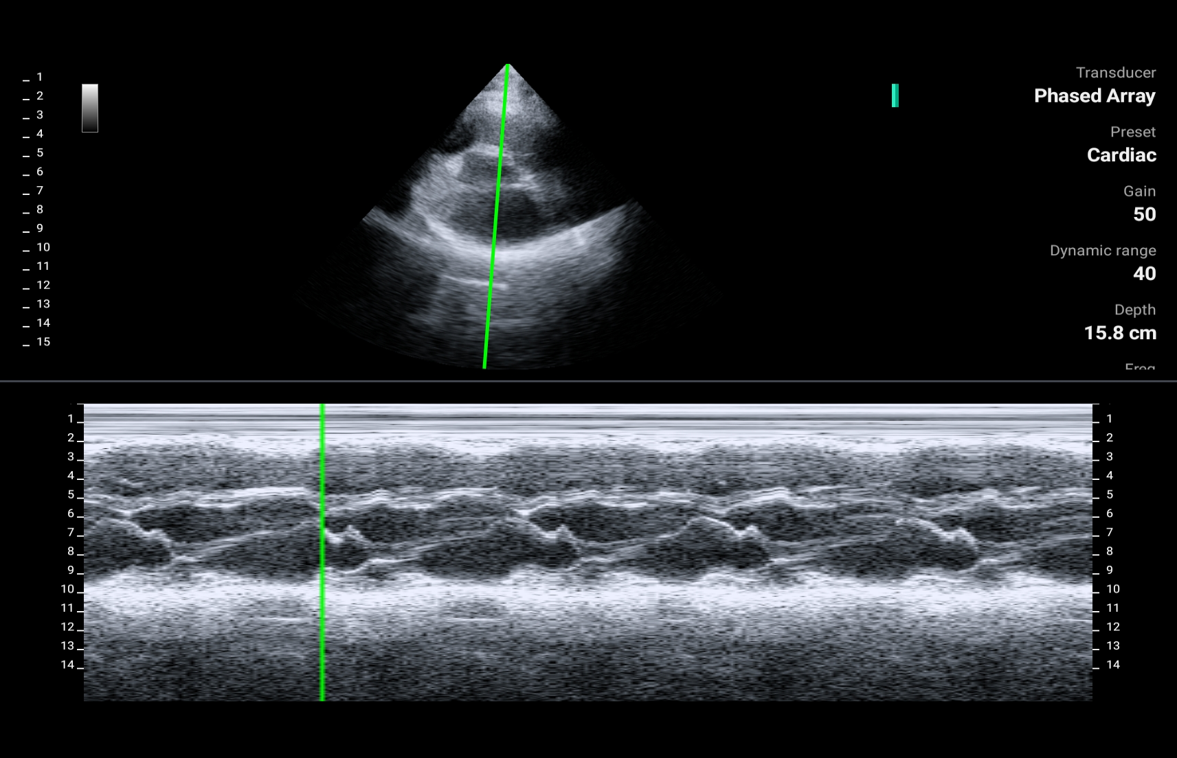
Parasternal long-axis view_M mode
-

Parasternal short-axis view_B mode
-
Parasternal short-axis view_B mode
-
.jpg)
Knee-patalla tendon(1)
-
Knee-patalla tendon
-
.png)
Median nerve_B mode(1)
-

Median nerve_B mode
-
_B mode.png)
Shoulder (Biceps tendon)_B mode
-
(1).png)
Shoulder(supraspinatus tendon)(1)
-
.png)
Shoulder(supraspinatus tendon)
-

OB Bmode
-
.png)
OB Bmode(1)
-
.png)
OB Bmode(2)
-
.png)
OB Bmode(3)
-

OB Brain Bmode
-
CCA Plaque_B mode
-
.jpg)
CCA Plaque_B mode(1)
-
.jpg)
CCA Plaque_B mode(2)
-
CCA B mode
-
CCA Color mode
-

CCA PW mode
-
Finger PD mode
-

Thyroid B mode
-
.png)
Thyroid B mode(1)
-
.png)
Thyroid B mode(2)
-
.png)
Thyroid B mode(3)
-
Uterus Bmode(Transvaginal sonography, TVS)
-
.png)
Uterus Bmode(Transvaginal sonography, TVS)(1)
-
.png)
Uterus Bmode(Transvaginal sonography, TVS)(2)
-
.png)
Uterus Bmode(Transvaginal sonography, TVS)(3)
-
.png)
Uterus Bmode(Transvaginal sonography, TVS)(4)
-
.png)
Uterus Bmode(Transvaginal sonography, TVS)(5)
-
Uterus Bmode(Transvaginal sonography, TVS)(6)
-
.png)
Uterus Bmode(Transvaginal sonography, TVS)(7)
-

Uterus Bmode(Transvaginal sonography, TVS)png
-
Uterus Ovary Bmode(Transvaginal sonography, TVS)
.png)
Helping to bring medical care to even the most remote locations
-

In-ambulance use
Rapid assessment of critical conditions, diagnosis of internal injuries of bleeding.
-

Emergency room
Performs the Focused Assessment with Sonography for Trauma (FAST) scans.
-

Telemedicine
Provides healthcare professionals with the ability to assess and diagnose patients remotely.
-

Clinicians on regular rounds
Enhances clinical assessments, improves patient care and makes informed decisions at the point of care.
-

Medical training
Strengths the learning experience across various medical disciplines.

ASUS Handheld Ultrasound LU800 Series
LU800L
| Types of array | Linear | Convex | Microconvex | Phased Array | Endocavity |
| Frequency | 4.2-12.5MHz | 2-5MHz | 3.6-8.5MHz | 1.3-3.7MHz | 3.6-8.5MHz |
| Max depth | 12.6 cm | 30 cm | 12.6 cm | 30 cm | 15 cm |
| Elements | 128 | 128 | 128 | 64 | 128 |
| Field of view | NA | 60° | 100° | 90° | 151° |
| Radius | NA | 60 mm | 15 mm | NA | 10 mm |
| Dimensions | 168 x 58 x 34 | 177 × 71 × 34 | 176 x 58 x 34 | 174 x 58 x 34 | 345 x 58 x 34 |
| Weight | 270 g | 305 g | 265 g | 280 g | 295 g |
| Image Modes | B / M / CF* / PW* / PD* | ||||
| Image file | JPEG / PNG / BMP / DICOM* | ||||
| Compatibility | ASUS MediConnect | ||||
| Battery | 3000mAh | ||||
| Intended use & Application | |||||
| Abdomen | |||||
| Abdomen difficult | |||||
| Renal | |||||
| GYN | |||||
| OB Mid Late | |||||
| Bladder Meas | |||||
| Peripheral vessels | |||||
| Thyroid | |||||
| Breast | |||||
| Superficial | |||||
| Musculoskeletal | |||||
| Carotid | |||||
| Cardiac | |||||
| Cardiac | |||||

| Types of array | Linear |
| Frequency | 4.2-12.5MHz |
| Max depth | 12.6 cm |
| Elements | 128 |
| Field of view | NA |
| Radius | NA |
| Dimensions | 168 x 58 x 34 |
| Weight | 305 g |
| Image Modes | B / M / CF* / PW* / PD* |
| Image file | JPEG / PNG / BMP / DICOM* |
| Compatibility | ASUS MediConnect |
| Battery | 3000mAh |
| Intended use &Application | |
| Peripheral vessels | |
| Thyroid | |
| Breast | |
| Superficial | |
| Musculoskeletal | |
| Carotid | |
| Types of array | Convex |
| Frequency | 2-5MHz |
| Max depth | 30 cm |
| Elements | 128 |
| Field of view | 60° |
| Radius | 60 mm |
| Dimensions | 177 × 71 × 34 |
| Weight | 270 g |
| Image Modes | B / M / CF* / PW* / PD* |
| Image file | JPEG / PNG / BMP / DICOM* |
| Compatibility | ASUS MediConnect |
| Battery | 3000mAh |
| Intended use &Application | |
| Abdomen | |
| Abdomen difficult | |
| Renal | |
| GYN | |
| OB Mid Late | |
| Bladder Meas | |
| Cardiac | |
| Types of array | Microconvex |
| Frequency | 3.6-8.5MHz |
| Max depth | 12.6 cm |
| Elements | 128 |
| Field of view | 100° |
| Radius | 15 mm |
| Dimensions | 176 x 58 x 34 |
| Weight | 265 g |
| Image Modes | B / M / CF* / PW* / PD* |
| Image file | JPEG / PNG / BMP / DICOM* |
| Compatibility | ASUS MediConnect |
| Battery | 3000mAh |
| Intended use &Application | |
| Abdomen | |
| Abdomen difficult | |
| Types of array | Phased Array |
| Frequency | 1.7-3.7MHz |
| Max depth | 30 cm |
| Elements | 64 |
| Field of view | 90° |
| Radius | NA |
| Dimensions | 174 x 58 x 34 |
| Weight | 280 g |
| Image Modes | B / M / CF* / PW* / PD* |
| Image file | JPEG / PNG / BMP / DICOM* |
| Compatibility | ASUS MediConnect |
| Battery | 3000mAh |
| Intended use &Application | |
| Abdomen | |
| Cardiac | |
| Types of array | Endocavity |
| Frequency | 3.6-8.5MHz |
| Max depth | 15 cm |
| Elements | 128 |
| Field of view | 151° |
| Radius | 10 mm |
| Dimensions | 345 x 58 x 34 |
| Weight | 295 g |
| Image Modes | B / M / CF* / PW* / PD* |
| Image file | JPEG / PNG / BMP / DICOM* |
| Compatibility | ASUS MediConnect |
| Battery | 3000mAh |
| Intended use &Application | |
| Abdomen | |
| Abdomen difficult | |
*Optional
**Availability depends on different regions.
ASUS is looking for ultrasound distributors
Contact us today to explore the opportunities and be part of our success story.
ASUS Handheld Ultrasound Glossary
M mode measurement
Ventricle Measure
- LVIDd : Left ventricular internal diameter end diastole
- LVIDs: Left ventricular internal diameter end systole
- EDV: End diastolic volume, amount of blood in the ventricle during diastole
- ESV: End systolic volume, amount of blood in ventricle after systole
- EDV Index: End-diastolic volume index, referred to as EDV corrected for BSA
= EDV / BSA
- ESV Index: End- systolic volume index
= EDV / BSA
- SV: Stroke volume, volume of blood pumped from the heart in one cycle of diastole and systole, is affected by Preload, contractility and Afterload
= EDV – ESV
- CO: Cardiac output, quantity of blood pumped per minute through the aorta and into the peripheral circulation, is proportional to (Arterial pressure / Total peripheral resistance)
= SV * HR
- EF: Ejection fraction, reflects the percentage of blood ejected from the ventricle
= SV / EDV * 100%
- FS: Fractional shortening
= (LVIDd – LVIDs) / LVIDd * 100%%
OB calculations
- GA: Gestational Age.
- Fetal age: Conceptional age.
- EFW: Estimated fetal weight.
- AUA: Arithmetic ultrasound age.
- DOC: Date of conception.
- LMP: Last menstrual period.
- EDD: Estimated due date.
In Early OB
- CRL: Crown-rump length
- GS: Gestational sac.
In Mid-late OB
- AC: Abdominal circumference.
- HC: Head circumference.
- FL: Femur length.
- BPD: Biparietal diameter.
References
- Early OB calculations: Hadlock 1992. Fetal Crown-Rump Length: Reevaluation of Relation to Menstrual Age with High-Resolution Real-Time US.
- Mid-late OB calculations: Hadlock 1984 Hadlock F.P, Deter R.L, Harrist R.B. and Park S.K, Estimating fetal age: computer-assisted analysis of multiple fetal growth parameters, Radiology, 152, pp 497-501, 1984
- EFW equations: EFW by AC, BPD FL, and HC: Hadlock 1985 Hadlock F.P, Harrist R.B, Sharman R.S, Deter R.L, Park S.K, Estimation of fetal weight with the use of head, body, and femur measurements–a prospective study, Am.J.Obstet.Gynecol., 151, pp 333-337, 1985
PW mode measurement
The glossary of "Auto Measure"
- HR (bpm):Heart rate
- PSV (cm/s):Peak systolic velocity
- EDV (cm/s):End diastolic velocity
- PI:Pulsatility index (of Gosling)
PI = (PSV–EDV)/MV
MV (cm/s):Mean velocity - RI:Resistance index (of Pourcelot)
RI = (PSV–EDV)/PSV - VTI (cm):Velocity-time integral
- TAV Max (cm/s):The maximum of time-averaged velocity
- S/B:The average RI of a cycle
- SD:Systolic/Diastolic Ratio
SD = PSV/EDV - ACCL (cm/s²):The acceleration index
ACCL = (PSV – EDV)/ACCT - ACCT (s):The time from the lowest (EDV) to the highest (PSV)
- VFM (ml/min):Volume flow per minute
- VFC (ml):Volume flow per cycle
- VFM Max (ml/min):The maximum of volume flow per minute
- Diam (mm) :Diameter
- *VFM, VFM Max, Diam values will be shown when you are in the frozen state.
The glossary of measure tool "PW V, T, HR"
- T (s):Time
- HR (bpm):Heart rate
- Range (cm/s):The range of flow velocity
The glossary of measure tool "RI, S/D"
- PSV (cm/s):Peak systolic velocity
- EDV (cm/s):End diastolic velocity
- S/D:Systolic/Diastolic Ratio
SD = PSV/EDV - RI:Resistance index (of Poucelot)
RI = (PSV–EDV)/PSV
The glossary of measure tool "PI"
- PSV (cm/s):Peak systolic velocity
- D (cm/s):End diastolic velocity
- Area (cm²):Blood vessel cross-sectional area
- Diam (mm):Diameter
- PI:Pulsatility index (of Gosling)
PI = (PSV–EDV)/MV
MV (cm/s):Mean velocity - VFM Max (ml/min):The maximum of volume flow per minute
- TAV Max (cm/s):The maximum of time-averaged velocity
The glossary of measure tool "VTI"
- VTI (cm):Velocity-time integral
Cardiac measurement in B mode
- BSA: Body Surface Area
= (Height * Weight / 3600)^½ - SV (ml): Stroke volume
= EDV – ESV - SI (ml/m²): Stroke volume index
= SV / BSA - CO (l/min): Cardiac output
= SV * HR / 1000 - CI (l/m/m²): Cardiac Index
= CO / BSA - EF (%): Ejection fraction
= SV / EDV * 100%


