


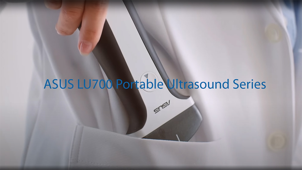
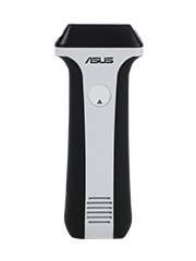
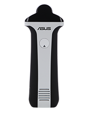
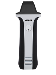
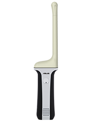
ASUS Handheld Ultrasound Solution LU700 / LU710
ASUS Handheld Ultrasound Solution
High-quality handheld scanners for fast, reliable medical imaging
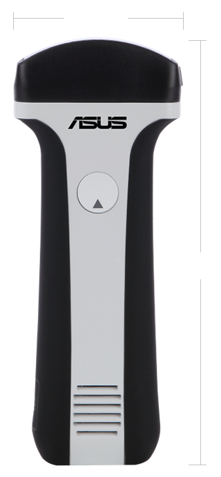
74 mm
187 mm
-
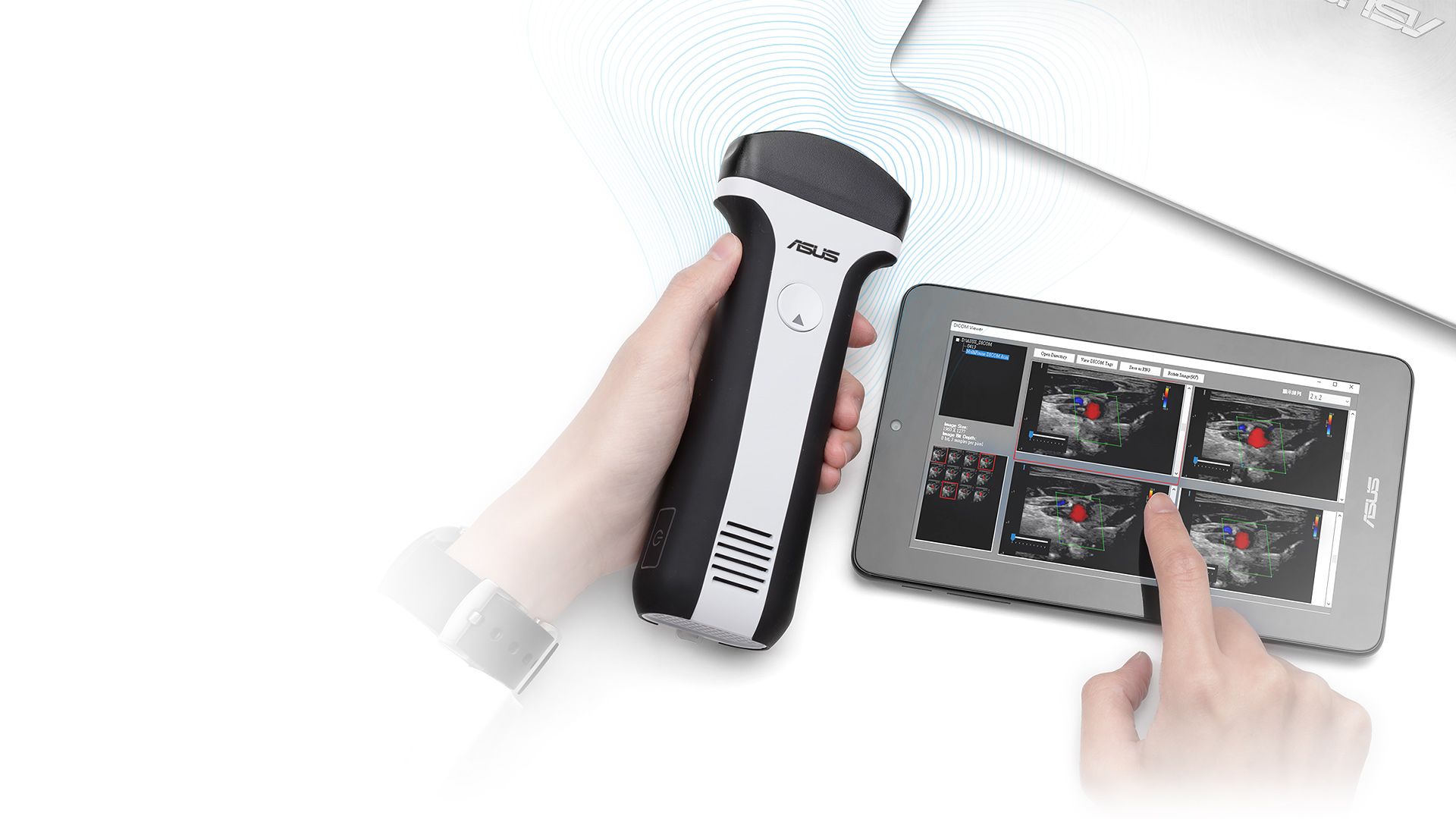
Handheld, smart imaging device
Lightweight, wireless handheld unit seamlessly integrates with proprietary imaging system software. -
Full functions and excellent image quality
Models produce high-quality imagery across diverse scenarios. -
Long battery life
Up to four hours of continuous use from a single charge. -
Intuitive interface and flexible image-management tools
Easy-to-use device interface and robust software* enable a range of uses by medical professionals
ASUS Handheld Ultrasound Series
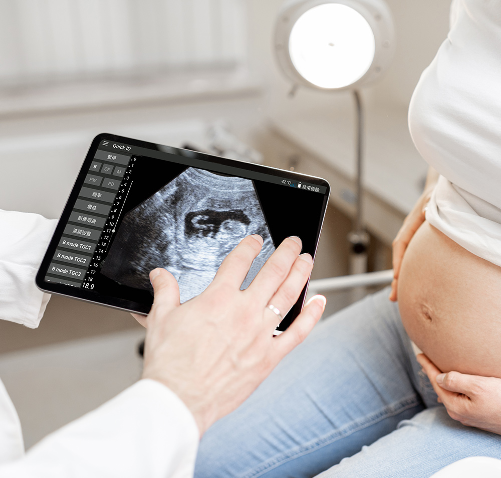
Clinic
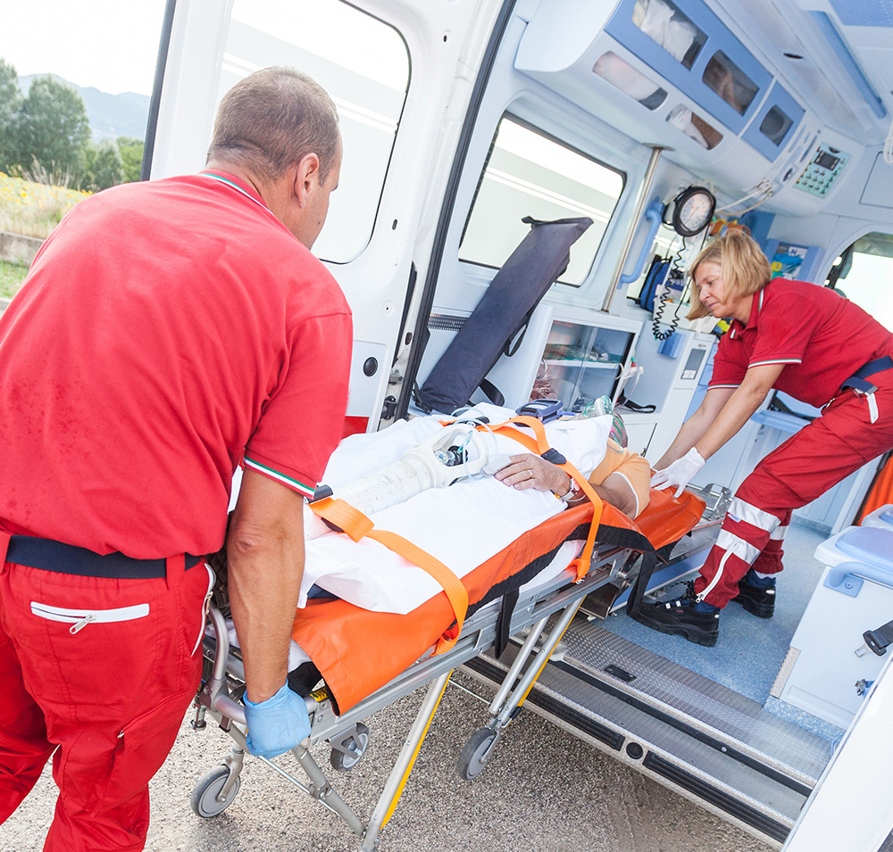
Ambulances

Clinicians on regular rounds

Emergency-room

Medical training

ASUS Handheld Ultrasound in Motion
-
Smart handheld scanners for fast, reliable medical imaging
The compact and wireless ASUS Handheld Ultrasound Series provides healthcare professionals with a comprehensive array of functions, exceptional scanned images, and energy efficiency. Click on the video to learn more about ASUS Handheld Ultrasound devices. -
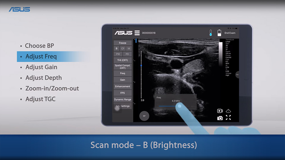
User Guide
Learn about the intuitive UI and extensive capabilities of ASUS Handheld Ultrasound Devices in this step-by-step video.
-
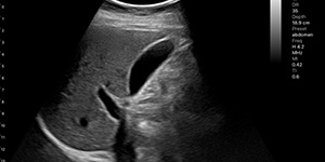
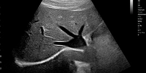
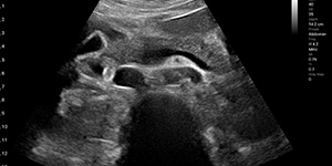
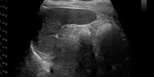
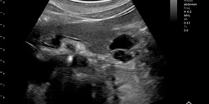
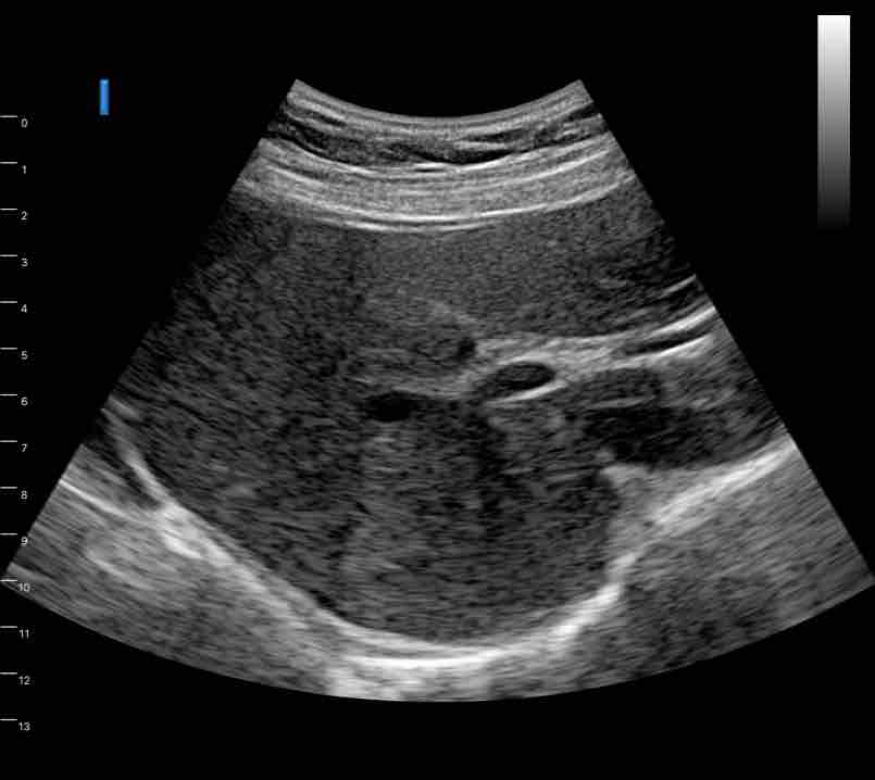
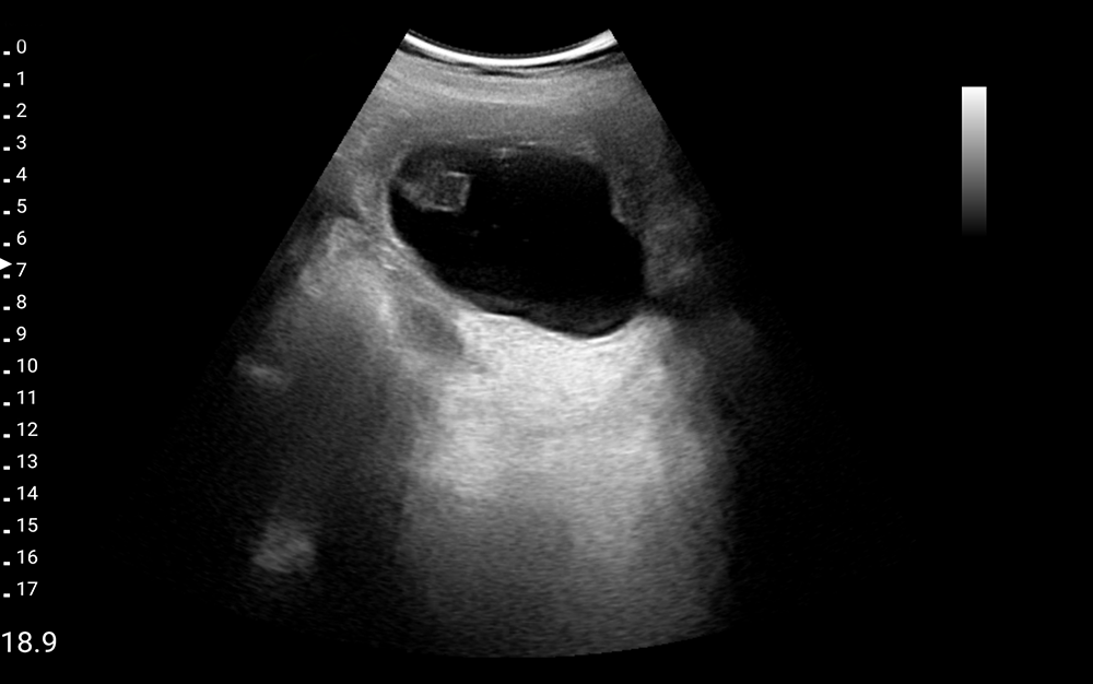
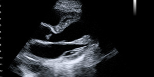
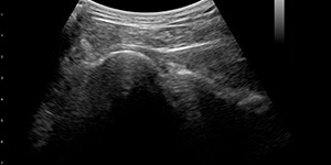
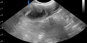
Gall bladder
-
Intraductal papillary mucinous neoplasm IPMN
-
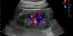
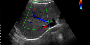
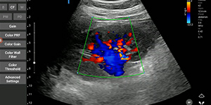
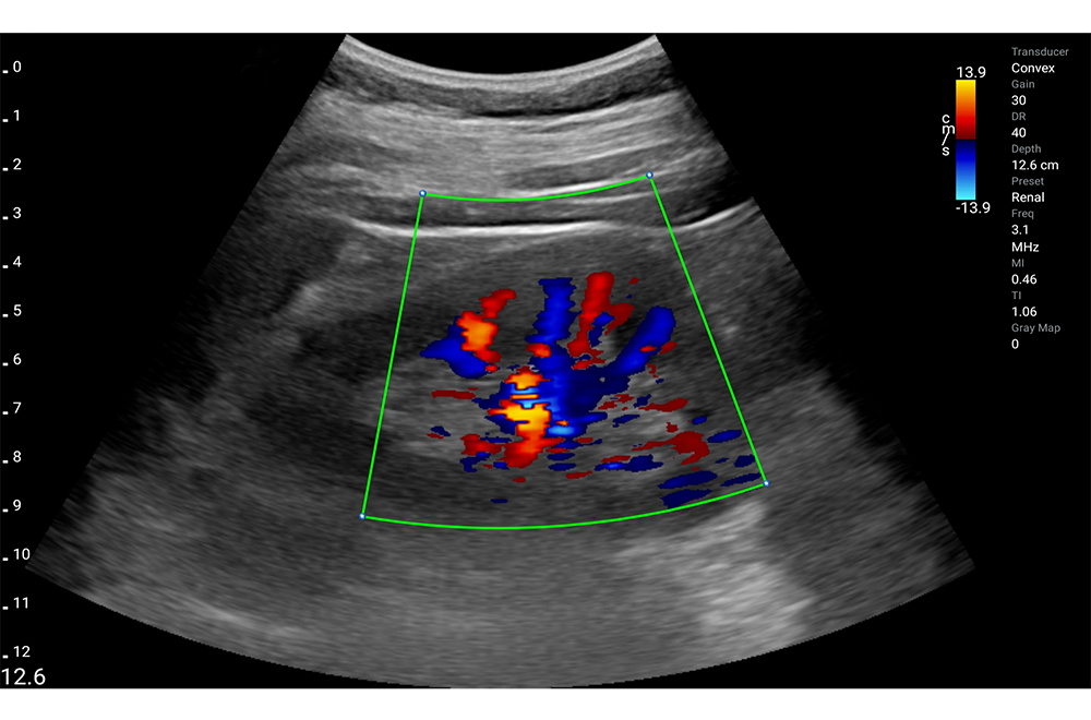
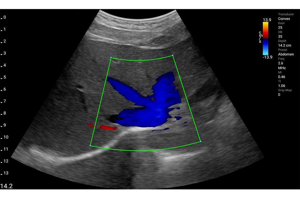
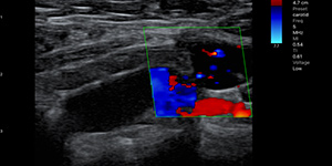
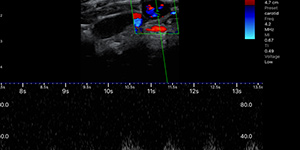
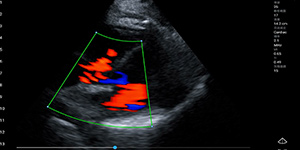
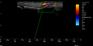
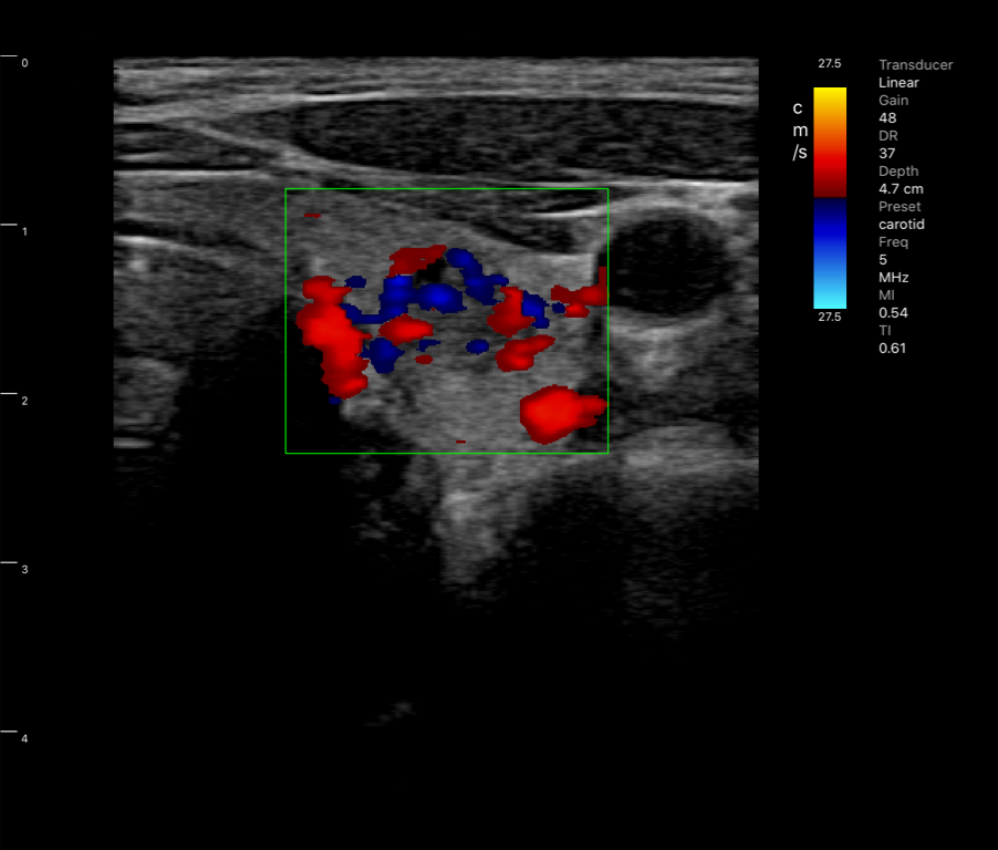
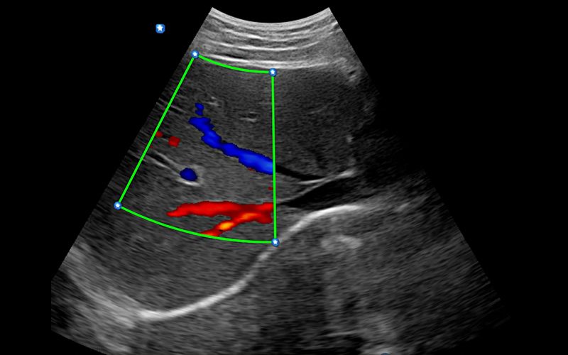
Kidney, Color doppler
-
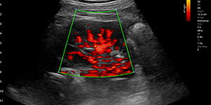
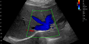
Kidney, Power doppler
-
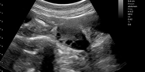
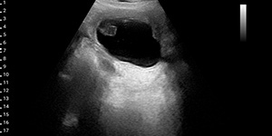
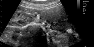
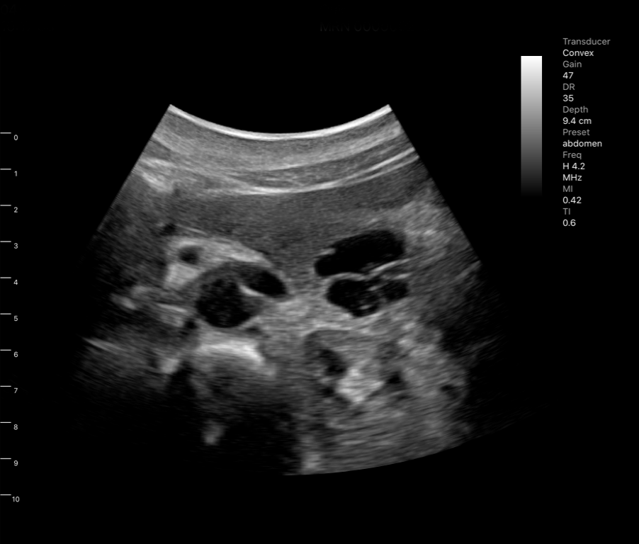
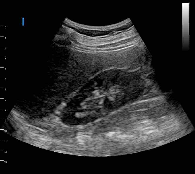
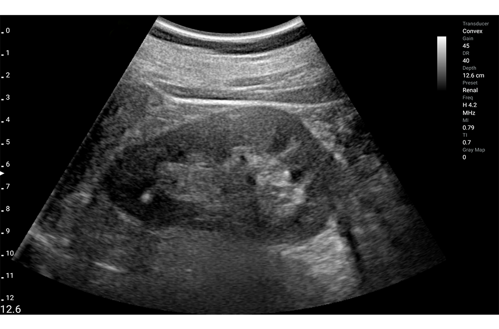
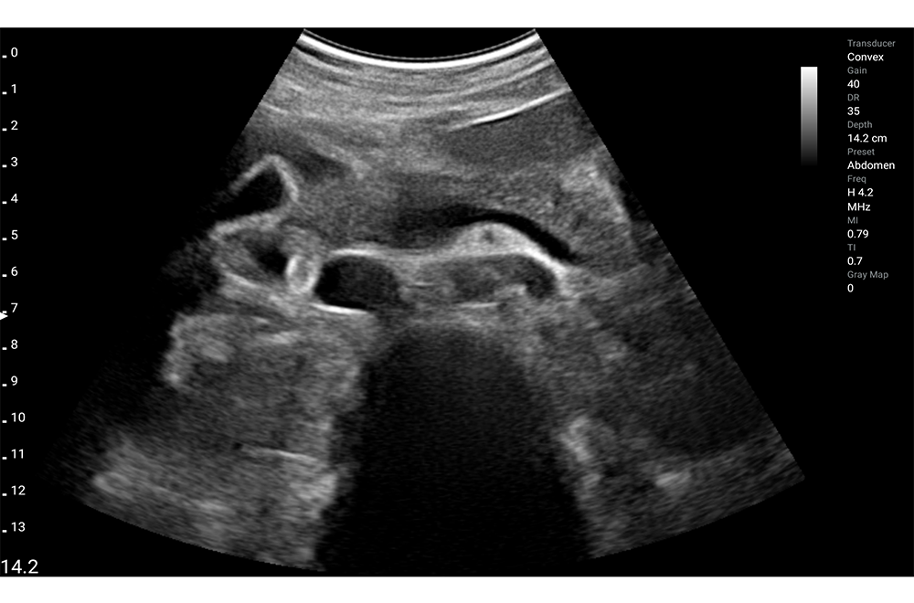
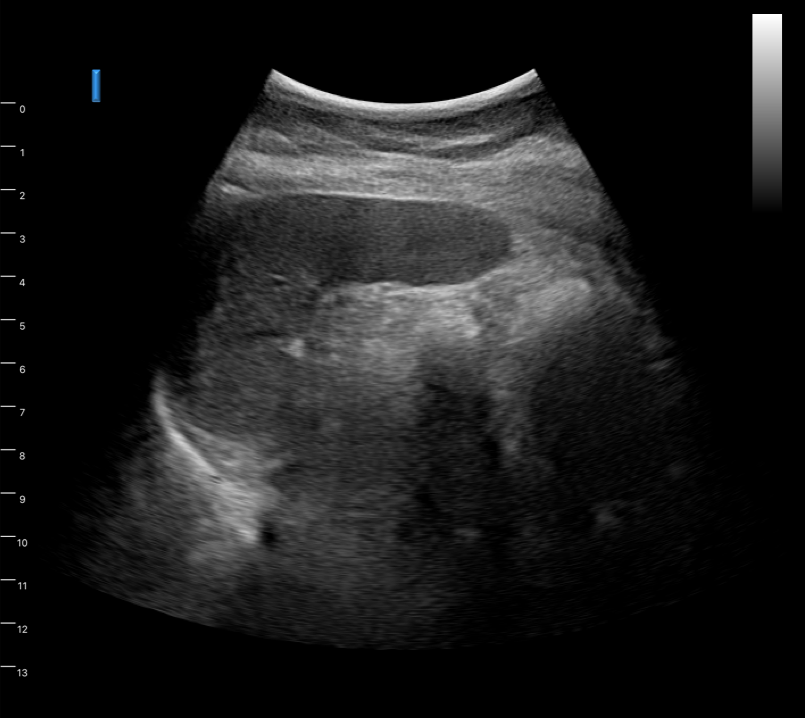
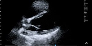
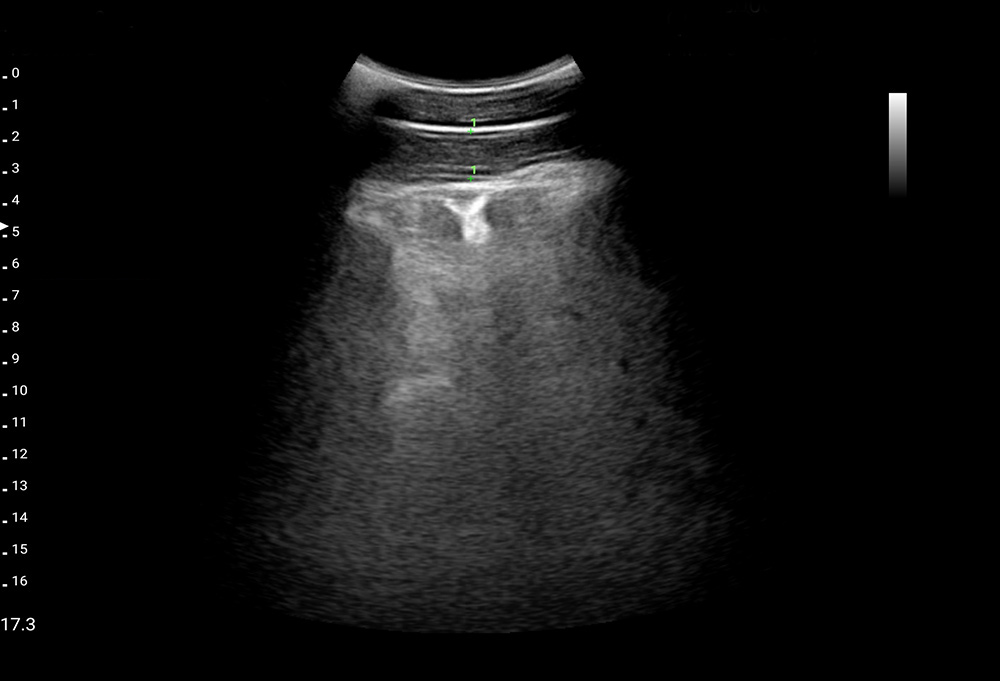
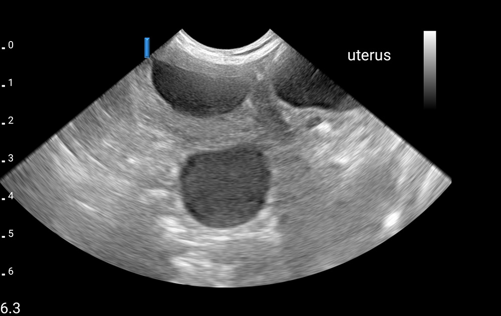
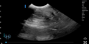
Kidney
-
Kidney
-
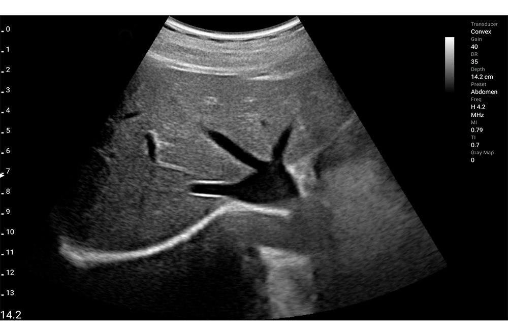
Liver tansverse
-
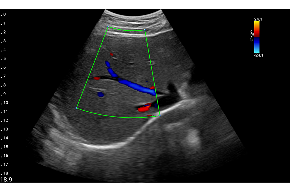
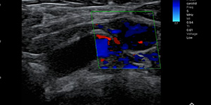
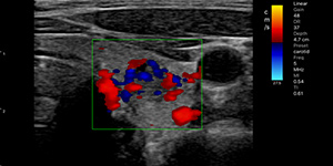
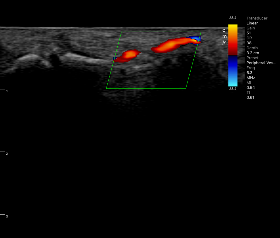
Liver, Color doppler
-
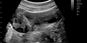
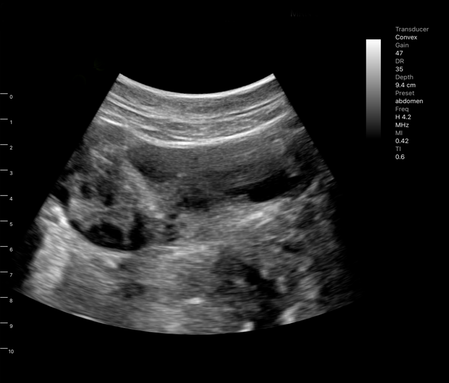
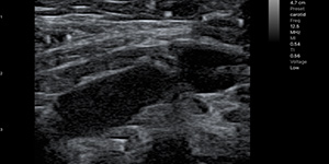
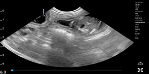
Liver
-
Lliver transverse, Color doppler
-
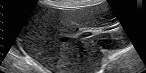
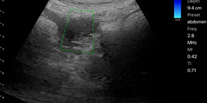
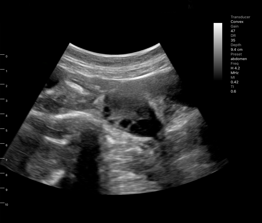
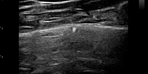
Pancreas
-
Polysystic ovary syndrome with follicles
-
Polysystic ovary syndrome with follicles2
-
Prostate gland
-
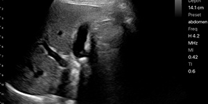
Spleen
-
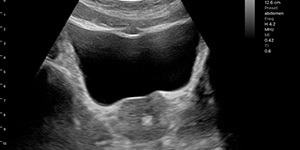
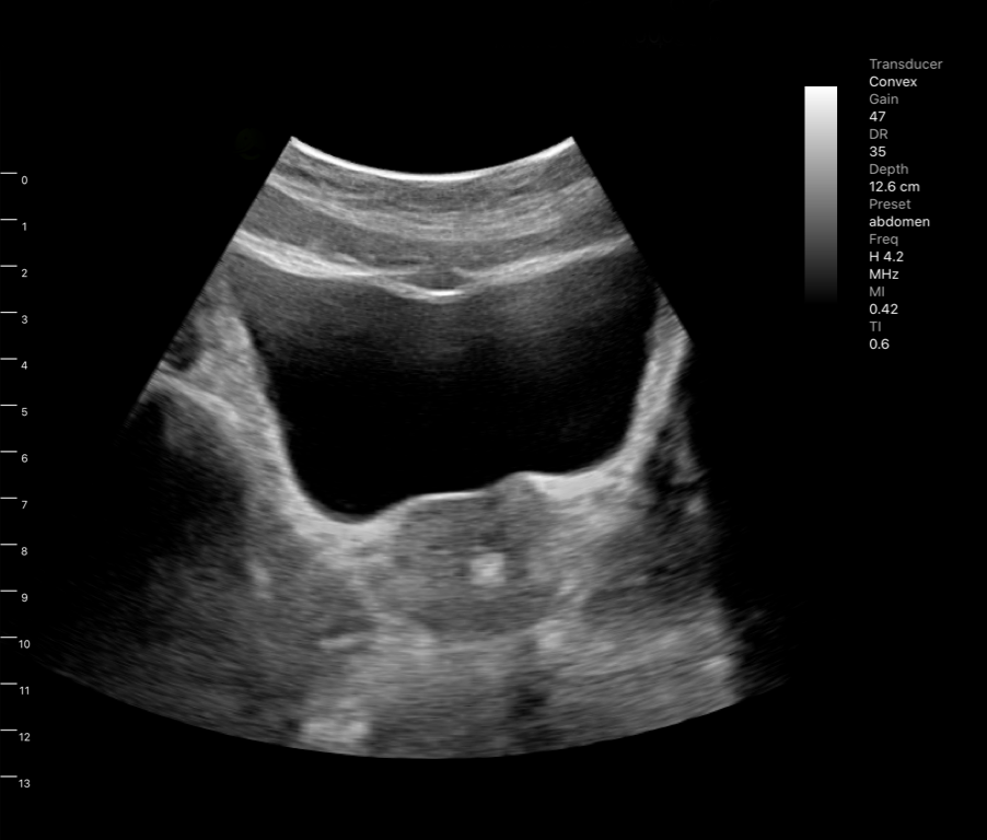
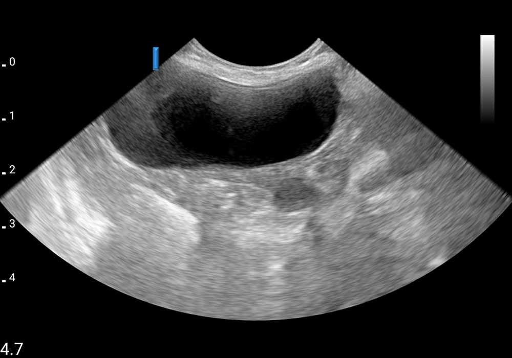
Urinary bladder mass
-
Colon tumor
-
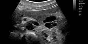
Gall bladder stones
-
Intraductal pancreatic mucinous neoplasm, IPMN
-
Kidney, Color doppler
-
Polycystic ovarian syndrome with follicles

more
close
-
Breast fibroadenoma
-
Post-vaccination axillary lymph node hyperplasia, color doppler
-
Post-vaccination axillary lymph node hyperplasia, pulse wave
-
Post-vaccination axillary lymph node hyperplasia
-
Normal lung HRUS
-
Post-vaccination axillary lymph node hyperplasia, Color doppler
-
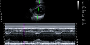
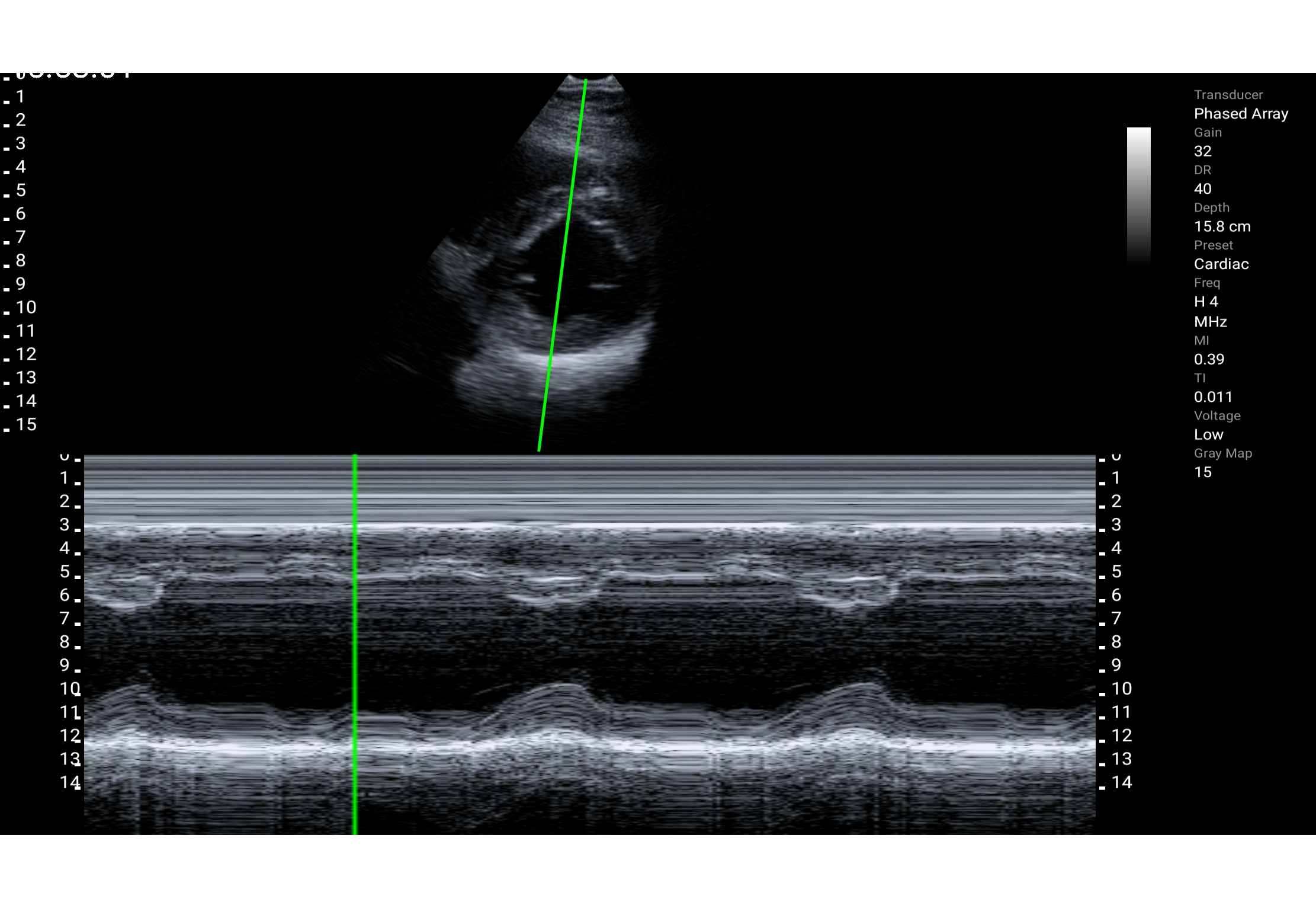
Parasternal short-axis view, M mode
-
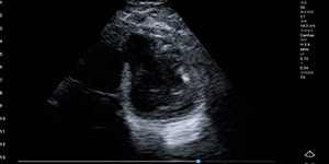
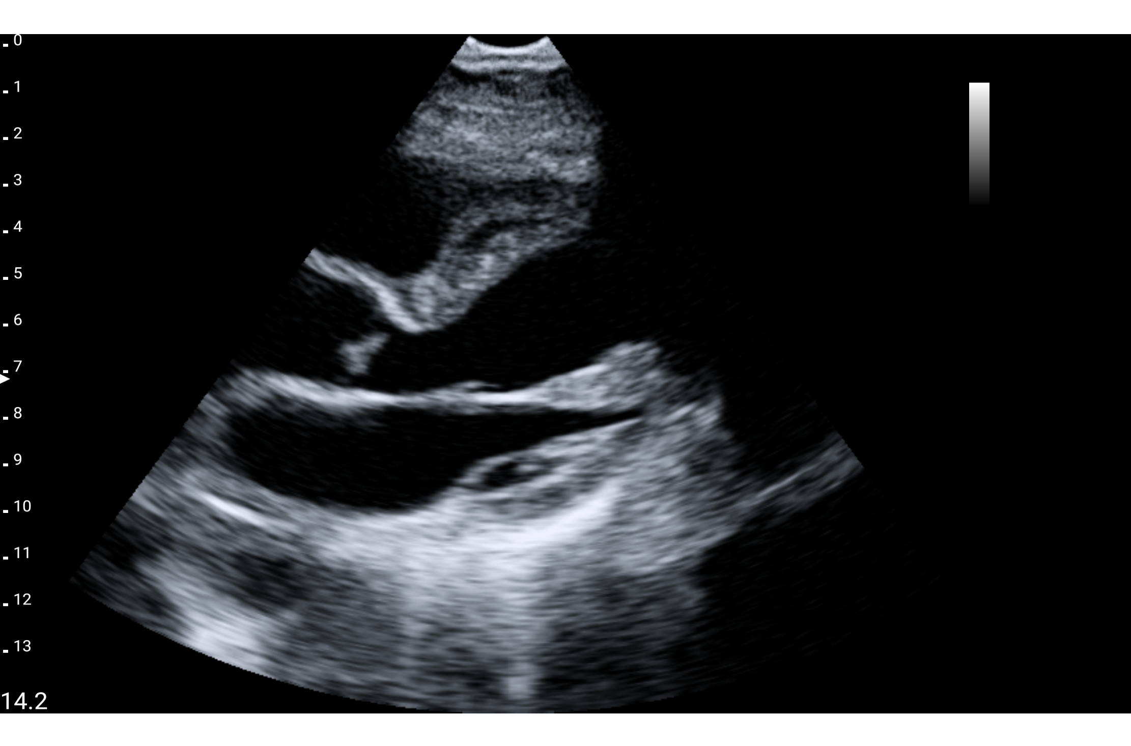
Parasternal long-axis view
-
Parasternal long-axis view
-
Parasternal short-axis view, Color doppler
-
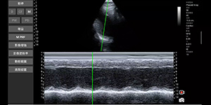
Parasternal short-axis view, M mode
-
Parasternal short-axis view
-
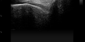
Humeral cartilage
-
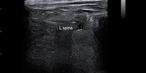
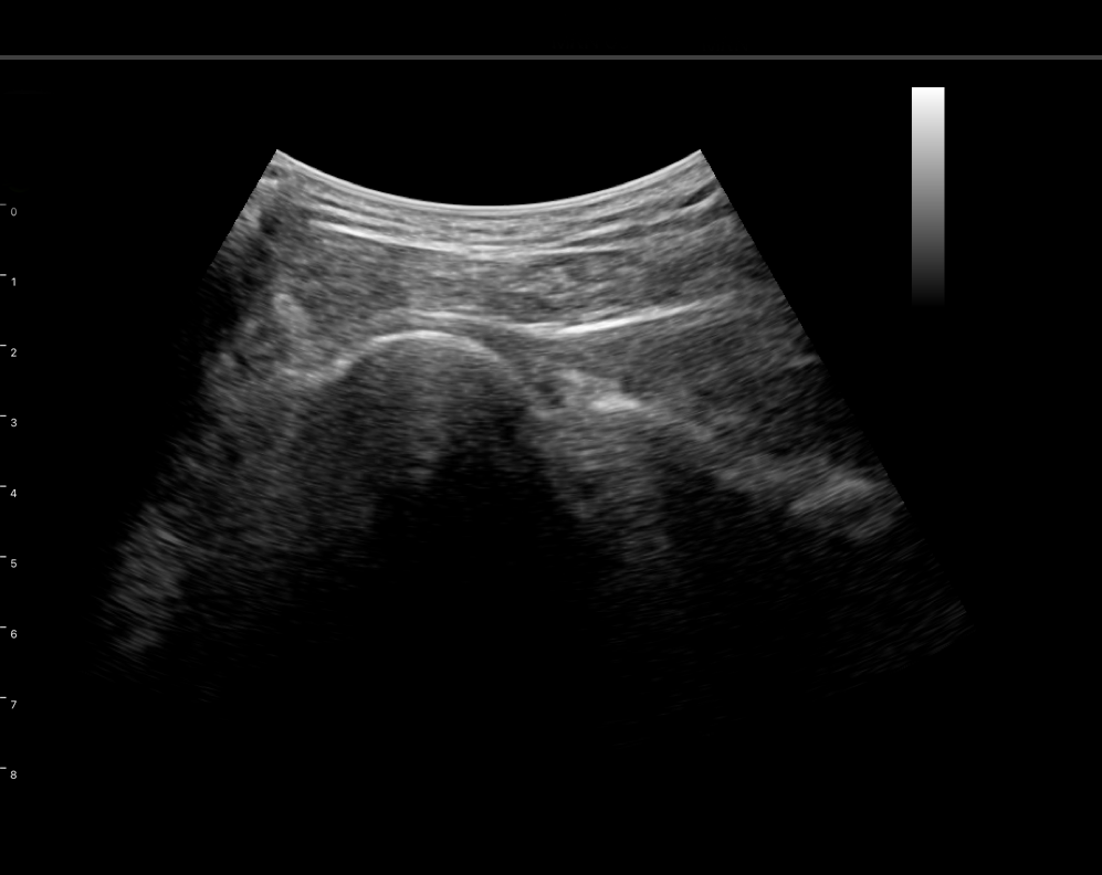
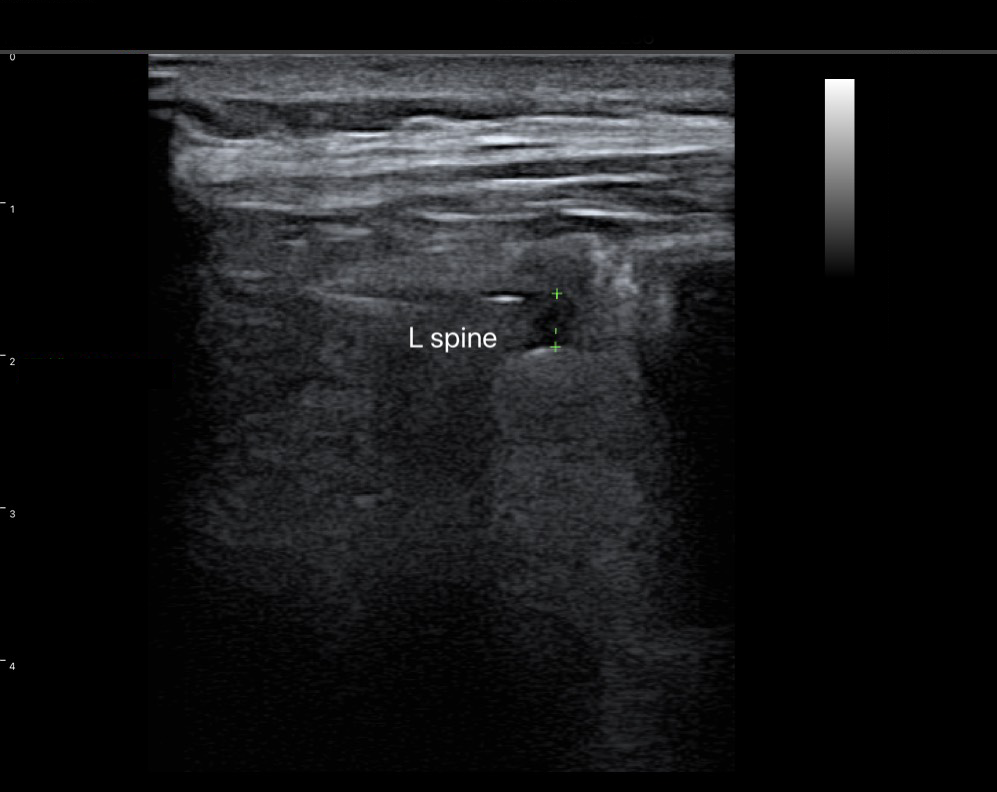
Vertebrae, L spine
-
Knee (DJD with femoral spur, buldging of medial meniscus)
-
Knee(ingrapatellar tendon)
-
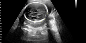
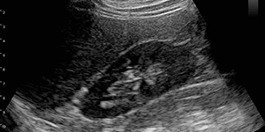
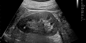
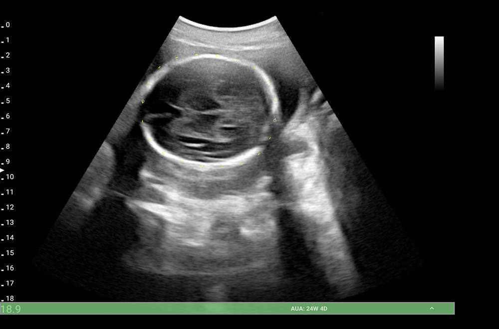
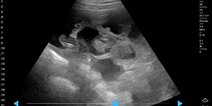
Fetus,-24wk
-
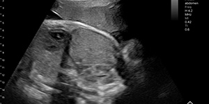
Fetus, 37wk
-
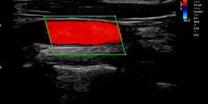
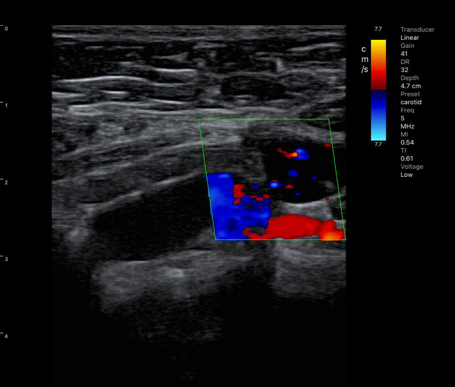
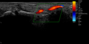
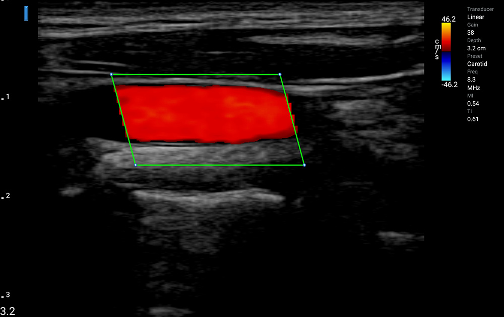
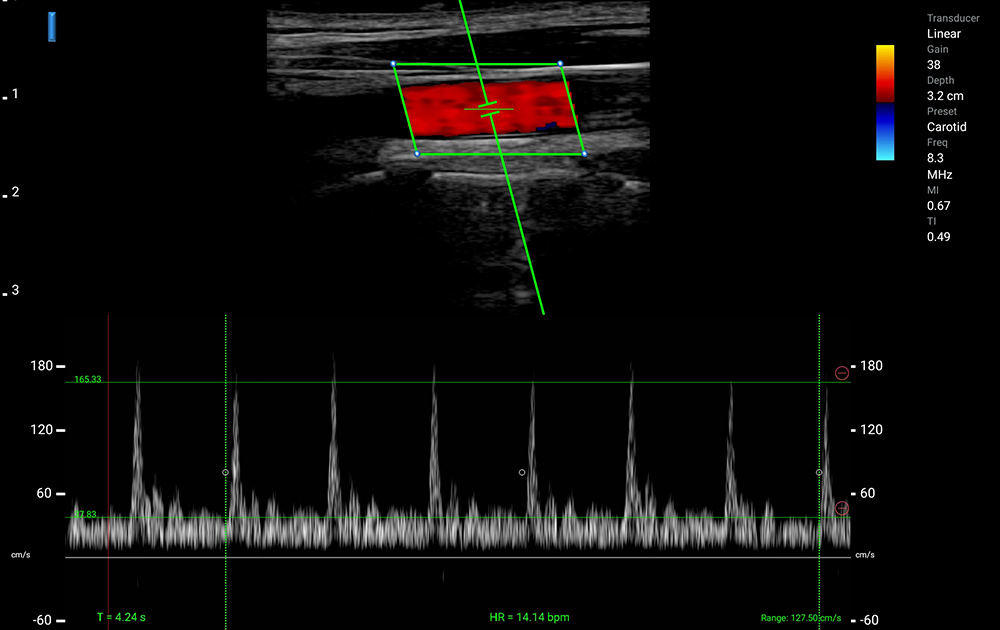
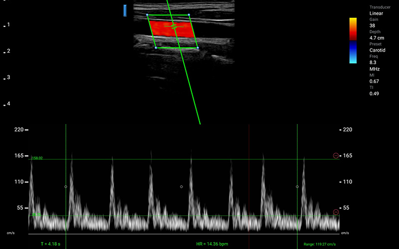
Carotid, Color doppler
-
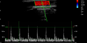
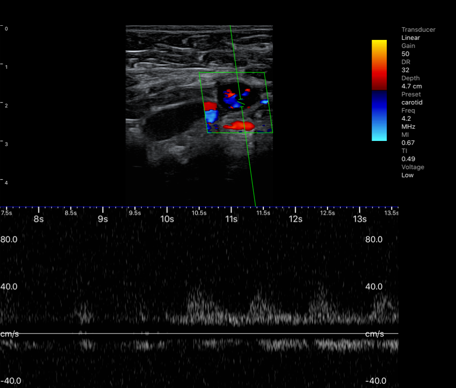
Carotid, Pulse wave
-
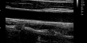
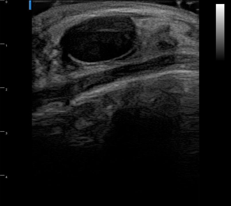
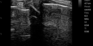
Carotid
-
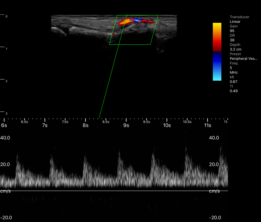
Finger peripheral v. Pulse wave
-
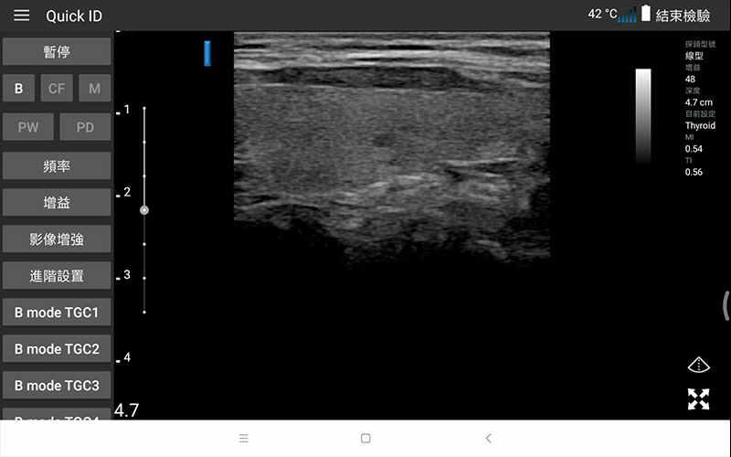
Thyroid gland
-
Finger peripheral v. Color doppler
-
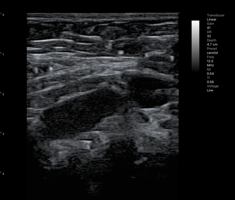
Carotid
-
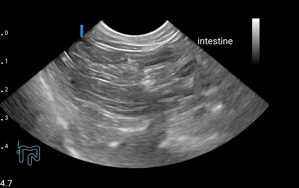
Esophagus
-
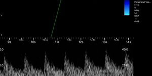
Finger peripheral vessel, Pulse wave
-
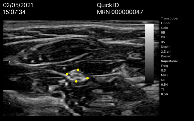
Thyroid gland cyst

-
Face NGT
-
Needle guided aspiration


ASUS Handheld Ultrasound for Medical Care
LU700C Convex
| Intended use | General abdominal imaging, lungs, muscles and bones (traditional), muscles and bones (surface), peripheral arteries, and obstetrics and gynecology. | General abdominal imaging, lungs, small organs (chest, thyroid), muscles and bones (traditional), muscles and bones (surface), and peripheral arteries. | General abdominal imaging, lungs, small organs (chest, thyroid), muscles and bones (traditional), muscles and bones (surface), and peripheral arteries. | General abdominal imaging, small organs, cardiac diagnosis, muscle and bone (traditional), muscle and bone (surface), and peripheral arteries. | Abdomen Cardiac | Endocavity |
| Frequency | 2.0 –5.0 MHz | 5.0 –10.0 MHz | 4.2 –12.5 MHz | 3.6 –8.5 MHz | 1.3-3.7MHz | 3.6-8.5MHz |
| FOV | 60° | N/A | N/A | 100° | 90° | 151° |
| Depth | 18 cm | 6 cm | 12.6 cm | 12.6 cm | 30 cm | 15 cm |
| Size | 187 x 74 x 40 mm | 178 x 74 x 40 mm | 178 x 74 x 40 mm | 190 x 74 x 40 mm | 194 × 74 × 40 mm | 370 x 74 x 40 mm |
| Weight | 388g | 357g | 357g | 340g | 350g | 412g |
| Operation | B/M/CF/PW/PD | |||||
| Image file | JPEG/PNG/BMP/DICOM | |||||
| Battery | Rechargeable 6000 mAhLi-ion Battery, up to 4 hours. | |||||
| Intended use | General abdominal imaging, lungs, muscles and bones (traditional), muscles and bones (surface), peripheral arteries, and obstetrics and gynecology. |
| Frequency | 2.0 –5.0 MHz |
| FOV | 60° |
| Depth | 25 cm |
| Size | 187 x 74 x 40 mm |
| Weight | 388g |
| Operation | B/M/CF/PW/PD |
| Image file | JPEG/PNG/BMP/DICOM |
| Battery | Rechargeable 6000 mAhLi-ion Battery, up to 4 hours. |
| Intended use | General abdominal imaging, lungs, small organs (chest, thyroid), muscles and bones (traditional), muscles and bones (surface), and peripheral arteries. |
| Frequency | 5.0 –10.0 MHz |
| FOV | N/A |
| Depth | 12 cm |
| Size | 178 x 74 x 40 mm |
| Weight | 357g |
| Operation | B/M/CF/PW/PD |
| Image file | JPEG/PNG/BMP/DICOM |
| Battery | Rechargeable 6000 mAhLi-ion Battery, up to 4 hours. |
| Intended use | General abdominal imaging, lungs, small organs (chest, thyroid), muscles and bones (traditional), muscles and bones (surface), and peripheral arteries. |
| Frequency | 4.5 - 12.5 MHz |
| FOV | N/A |
| Depth | 12 cm |
| Size | 178 x 74 x 40 mm |
| Weight | 357g |
| Operation | B/M/CF/PW/PD |
| Image file | JPEG/PNG/BMP/DICOM |
| Battery | Rechargeable 6000 mAhLi-ion Battery, up to 4 hours. |
| Intended use | General abdominal imaging, small organs, cardiac diagnosis, muscle and bone (traditional), muscle and bone (surface), and peripheral arteries. |
| Frequency | 4.0 –8.0 MHz |
| FOV | 100° |
| Depth | 12 cm |
| Size | 190x 74 x 40 mm |
| Weight | 340g |
| Operation | B/M/CF/PW/PD |
| Image file | JPEG/PNG/BMP/DICOM |
| Battery | Rechargeable 6000 mAhLi-ion Battery, up to 4 hours. |
| Intended use | Abdomen Cardiac |
| Frequency | 1.7-3.7MHz |
| FOV | 90° |
| Depth | 18 cm |
| Size | 194 × 74 × 40 mm |
| Weight | 350g |
| Operation | B/M/CF/PW/PD |
| Image file | JPEG/PNG/BMP/DICOM |
| Battery | Rechargeable 6000 mAhLi-ion Battery, up to 4 hours. |
| Intended use | Endocavity |
| Frequency | 4-8.5MHz |
| FOV | 151° |
| Depth | 15 cm |
| Size | 370 x 74 x 40 mm |
| Weight | 412g |
| Operation | B/M/CF/PW/PD |
| Image file | JPEG/PNG/BMP/DICOM |
| Battery | Rechargeable 6000 mAhLi-ion Battery, up to 4 hours. |
* Availability depends on different regions.
Tutorial Video
-
Download
-
Connect the probe with app
-
Improving the quality of Wi-Fi connection
-
Review exams without connecting the probe
-
Download worklist
-
Edit patient information
-
Review exams with connecting the probe
-
Batch management and export images and videos
-
Export report

-
Export images to DICOM server
-
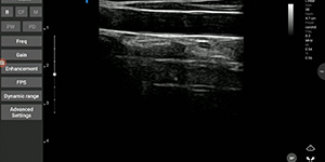
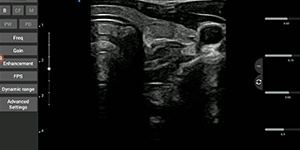
Adjusting depth
-
Adjusting gain
-
Adjusting freqency

-
Changing image orientation
-
Adjusting freeze timer
-
Using zoom
-
Capturing images in instant

-
Capturing cineloops in instant

-
Changing and customizing the preset

-
Adding annotations
-
Using body mark

-
Using measurement
-
Measuring the bladder volume
-
Comparing the images with dual screen
-
Calculating the gestational age
-
Measuring in OB early
-
Measuring in OB mid late

-
Saving images

-
Making videos
-
End exam
-
Adjusting TGC in B mode
-
Adjusting focus in B mode
-
Controlling M mode
-
The measurement in M mode
-
Adjusting ROI in C-PD mode
-
Controlling Pre-PW mode
-
Controlling PW mode
-
The measurement in PW mode
ASUS Handheld Ultrasound Glossary
M mode measurement
Ventricle Measure
- LVIDd : Left ventricular internal diameter end diastole
- LVIDs: Left ventricular internal diameter end systole
- EDV: End diastolic volume, amount of blood in the ventricle during diastole
- ESV: End systolic volume, amount of blood in ventricle after systole
- EDV Index: End-diastolic volume index, referred to as EDV corrected for BSA
= EDV / BSA
- ESV Index: End- systolic volume index
= EDV / BSA
- SV: Stroke volume, volume of blood pumped from the heart in one cycle of diastole and systole, is affected by Preload, contractility and Afterload
= EDV – ESV
- CO: Cardiac output, quantity of blood pumped per minute through the aorta and into the peripheral circulation, is proportional to (Arterial pressure / Total peripheral resistance)
= SV * HR
- EF: Ejection fraction, reflects the percentage of blood ejected from the ventricle
= SV / EDV * 100%
- FS: Fractional shortening
= (LVIDd – LVIDs) / LVIDd * 100%%
OB calculations
- GA: Gestational Age.
- Fetal age: Conceptional age.
- EFW: Estimated fetal weight.
- AUA: Arithmetic ultrasound age.
- DOC: Date of conception.
- LMP: Last menstrual period.
- EDD: Estimated due date.
In Early OB
- CRL: Crown-rump length
- GS: Gestational sac.
In Mid-late OB
- AC: Abdominal circumference.
- HC: Head circumference.
- FL: Femur length.
- BPD: Biparietal diameter.
References
- Early OB calculations: Hadlock 1992. Fetal Crown-Rump Length: Reevaluation of Relation to Menstrual Age with High-Resolution Real-Time US.
- Mid-late OB calculations: Hadlock 1984 Hadlock F.P, Deter R.L, Harrist R.B. and Park S.K, Estimating fetal age: computer-assisted analysis of multiple fetal growth parameters, Radiology, 152, pp 497-501, 1984
- EFW equations: EFW by AC, BPD FL, and HC: Hadlock 1985 Hadlock F.P, Harrist R.B, Sharman R.S, Deter R.L, Park S.K, Estimation of fetal weight with the use of head, body, and femur measurements–a prospective study, Am.J.Obstet.Gynecol., 151, pp 333-337, 1985
PW mode measurement
The glossary of "Auto Measure"
- HR (bpm):Heart rate
- PSV (cm/s):Peak systolic velocity
- EDV (cm/s):End diastolic velocity
- PI:Pulsatility index (of Gosling)
PI = (PSV–EDV)/MV
MV (cm/s):Mean velocity - RI:Resistance index (of Pourcelot)
RI = (PSV–EDV)/PSV - VTI (cm):Velocity-time integral
- TAV Max (cm/s):The maximum of time-averaged velocity
- S/B:The average RI of a cycle
- SD:Systolic/Diastolic Ratio
SD = PSV/EDV - ACCL (cm/s²):The acceleration index
ACCL = (PSV – EDV)/ACCT - ACCT (s):The time from the lowest (EDV) to the highest (PSV)
- VFM (ml/min):Volume flow per minute
- VFC (ml):Volume flow per cycle
- VFM Max (ml/min):The maximum of volume flow per minute
- Diam (mm) :Diameter
- *VFM, VFM Max, Diam values will be shown when you are in the frozen state.
The glossary of measure tool "PW V, T, HR"
- T (s):Time
- HR (bpm):Heart rate
- Range (cm/s):The range of flow velocity
The glossary of measure tool "RI, S/D"
- PSV (cm/s):Peak systolic velocity
- EDV (cm/s):End diastolic velocity
- S/D:Systolic/Diastolic Ratio
SD = PSV/EDV - RI:Resistance index (of Poucelot)
RI = (PSV–EDV)/PSV
The glossary of measure tool "PI"
- PSV (cm/s):Peak systolic velocity
- D (cm/s):End diastolic velocity
- Area (cm²):Blood vessel cross-sectional area
- Diam (mm):Diameter
- PI:Pulsatility index (of Gosling)
PI = (PSV–EDV)/MV
MV (cm/s):Mean velocity - VFM Max (ml/min):The maximum of volume flow per minute
- TAV Max (cm/s):The maximum of time-averaged velocity
The glossary of measure tool "VTI"
- VTI (cm):Velocity-time integral
Cardiac measurement in B mode
- BSA: Body Surface Area
= (Height * Weight / 3600)^½ - SV (ml): Stroke volume
= EDV – ESV - SI (ml/m²): Stroke volume index
= SV / BSA - CO (l/min): Cardiac output
= SV * HR / 1000 - CI (l/m/m²): Cardiac Index
= CO / BSA - EF (%): Ejection fraction
= SV / EDV * 100%
ASUS is looking for ultrasound distributors.
Contact us today to explore the opportunities and be part of our success story.


